Images and videos
Images


Osteochondritis dissecans
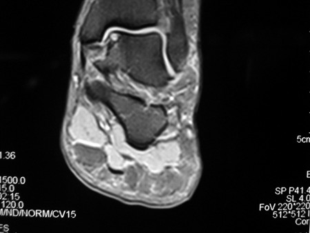
Coronal MRI image of the talus showing an osteochondral lesion on the medial aspect of the talar dome
Gupta RK, Kansay R, Aggarwal V, et al. Osteochondritis dessicans of the talus in a 26-year-old woman. BMJ Case Reports 2009; doi:10.1136/bcr.06.2008.0091
See this image in context in the following section/s:
Osteochondritis dissecans
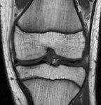
Osteochondral lesion of medial talus
From the collection of H. Chambers, MD
See this image in context in the following section/s:
Osteochondritis dissecans
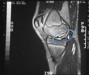
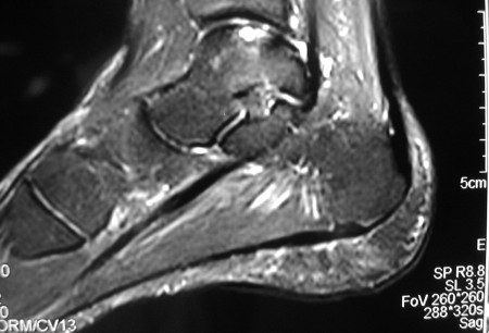
Sagittal magnetic resonance image (MRI) of the talus, showing an osteochondral lesion on the posterior aspect of the talar dome
Gupta RK, Kansay R, Aggarwal V, et al. Osteochondritis dessicans of the talus in a 26-year-old woman. BMJ Case Reports 2009; doi:10.1136/bcr.06.2008.0091
See this image in context in the following section/s:
Osteochondritis dissecans
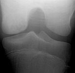
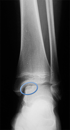
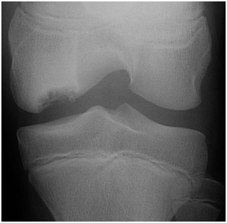
Tunnel view of osteochondritis dissecans
From the collection of H. Chambers, MD
See this image in context in the following section/s:

Osteochondritis dissecans
Healed osteochondritis dissecans
From the collection of H. Chambers, MD
See this image in context in the following section/s:

Osteochondritis dissecans
Healed osteochondritis dissecans
From the collection of H. Chambers, MD
See this image in context in the following section/s:
Osteochondritis dissecans
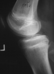
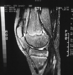
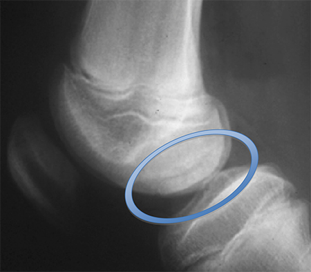
Preoperative radiograph with large osteochondritis lesion of the femoral condyle
From the collection of H. Chambers, MD
See this image in context in the following section/s:
Osteochondritis dissecans
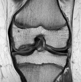
Preoperative anteroposterior magnetic resonance imaging (MRI)
From the collection of H. Chambers, MD
See this image in context in the following section/s:

Osteochondritis dissecans
Preoperative lateral magnetic resonance imaging (MRI) with no articular cartilage involvement
From the collection of H. Chambers, MD
See this image in context in the following section/s:

Osteochondritis dissecans
Magnetic resonance imaging (MRI) of knee demonstrating healing
From the collection of H. Chambers, MD
See this image in context in the following section/s:

Osteochondritis dissecans
After drilling of lesion, complete resolution (healing) of osteochondritis dissecans lesion
From the collection of H. Chambers, MD
See this image in context in the following section/s:
Osteochondritis dissecans
Coronal magnetic resonance image (MRI) of the talus showing an osteochondral lesion on the medial aspect of the talar dome
Gupta RK, Kansay R, Aggarwal V, et al. Osteochondritis dessicans of the talus in a 26-year-old woman. BMJ Case Reports 2009; doi:10.1136/bcr.06.2008.0091
See this image in context in the following section/s:

Osteochondritis dissecans
Healing after drilling of lesion
From the collection of H. Chambers, MD
See this image in context in the following section/s:
Use of this content is subject to our disclaimer