Tests
1st tests to order
plain radiographs
Test
Anterior soft-tissue swelling may be the only finding present early in the disease process.
Later in the course of the disease, findings may include an enlarged tibial tubercle, irregular ossification of the tubercle, fragmentation of the tubercle, or formation of a separate ossicle.[1][5][Figure caption and citation for the preceding image starts]: Knee x-ray in a patient with OSD (lateral view)From the collection of Dr J. Rosen [Citation ends].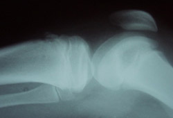
Should be ordered initially if symptoms are unilateral, severe, or persistent, and if there is any history of trauma.[1]
Result
may be normal; often demonstrate enlarged tibial tubercle, sometimes with fragmentation of the apophysis
Tests to consider
ultrasonography
Test
Performed if diagnosis uncertain.
Due to its ability to evaluate soft tissues and noncalcified cartilage, may provide insight into abnormal findings in these tissues early in the disease process.[5]
Result
may demonstrate pretibial swelling, fragmentation of the ossification center, thickening of the patellar tendon insertion, fluid accumulation in the infrapatellar bursa
MRI
Test
Performed if diagnosis uncertain.
Demonstrates pathologic findings early in the disease process. A variety of findings have been described.
MRI finding may be classified into 5 stages: normal, early, progressive, terminal, and healing.[14]
Early changes include edema at the tibial tubercle.
Progressive stage of the disease is characterized by partial avulsion of the secondary ossification center, usually pulled proximally.
At the terminal stage of the disease, complete separation of the avulsed secondary ossification center may be noted. Furthermore, there is swelling of the patellar tendon, indicating patellar tendonitis as a secondary pathologic change in OSD.
In the healing stage, the separated fragment is either ossified into a separate ossicle, or healed back to the apophysis via ossification of a fibrocartilaginous bridge that spanned the gap between the apophysis and the fragment.
Do not order MRI scans for children until plain radiographic examinations have been completed.[13]
Result
edema at the tibial tubercle, partial and/or complete separation of the fragments from the secondary ossification center, formation of a separate ossicle, or healing and remodeling of the apophysis
Use of this content is subject to our disclaimer