Tests
1st tests to order
clinical diagnosis
Test
High-altitude illness is primarily a clinical diagnosis, where patients present with typical findings in association with high-altitude travel.
Result
features of high-altitude illness
Tests to consider
arterial blood gases
Test
High-altitude pulmonary edema (HAPE) may demonstrate respiratory alkalosis.
Result
HAPE: reduced PaO2 and low to normal PaCO2
chest radiography
Test
The onset of radiographic changes in HAPE can be highly variable.[55] However, most cases eventually develop asymmetric areas of "cotton wool" infiltrates in the mid and lower zones of the lung fields.[56][Figure caption and citation for the preceding image starts]: CXR of high-altitude pulmonary edemaPublished with the kind permission of the Wilderness Medical Society [Citation ends].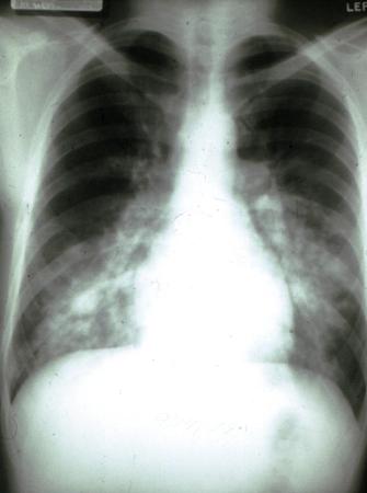
Changes tend to begin in the right mid zone and eventually spread across to the left. The apices and costophrenic angles are usually spared.
Although prominent pulmonary vasculature may be present, this is a common finding in anyone ascending to altitude.[58]
Signs of cardiogenic pulmonary edema are usually absent. Complete resolution of pulmonary infiltrates occurs quickly following recovery.[59]
Result
HAPE: asymmetric areas of "cotton wool" infiltrates in the mid and lower zones of the lung fields
ECG
Test
In HAPE, the ECG usually shows a sinus tachycardia and changes compatible with acute pulmonary hypertension. These include right axis deviation and bundle branch block; peaked P waves in leads II, III, and aVF; and an increase in the depth of precordial S waves.[55]
Result
HAPE: sinus tachycardia and changes compatible with acute pulmonary hypertension
chest ultrasound and echocardiography
Test
In HAPE, the presence of edema can result in the formation of "comet-tail artifacts" that are visible on ultrasound scanning. These, when assessed over 28 separate lung fields, can provide an objective assessment of pulmonary edema and can be used to monitor the course of the disease.[57][60]
The presence of a patent foramen ovale (present in 25% of the general population) may be associated with an increasing susceptibility to HAPE.[61]
Result
HAPE: "comet-tail artifacts"
WBC count
Test
The majority of laboratory investigations are normal in high-altitude illness.
In some cases of HAPE a small elevation in the WBC count occurs; this is due to a mild neutrophil leukocytosis.[55]
Result
elevated
lumbar puncture
Test
An increase in intracranial pressure is often seen in high-altitude cerebral edema (HACE). In advanced cases of HACE this can exceed normal values by up to 300 mm H2O.[62]
Cerebrospinal fluid (CSF) analysis may reveal the presence of red blood cells in severe cases, but in the vast majority CSF will be normal.[4]
Result
HACE: elevated intracranial pressure, RBCs
CT head
Test
In keeping with an increase in intracranial pressure CT scanning can reveal compression of the ventricles and changes to the gyri and sulci present on the surface of the cerebral hemispheres.
Changes seen on CT may take weeks or even months to resolve after clinical recovery.[14][23]
Result
compression of ventricles
MRI head
Test
MRI scanning shows formation of edema in the white matter. This is often concentrated in the splenium of the corpus callosum. Gray matter is largely unaffected by high-altitude cerebral edema. Changes seen on MRI may take weeks or even months to resolve after clinical recovery.[14][23]
Result
edema in white matter
Use of this content is subject to our disclaimer