Approach
The appearance of new symptoms at altitude should be assumed to be due to high-altitude illness until proved otherwise. An accurate history and an appropriate physical exam are often all that is required to diagnose acute mountain sickness (AMS), high-altitude pulmonary edema (HAPE), and high-altitude cerebral edema (HACE) in the field. Anyone with AMS presenting with abnormal neurologic findings must be assumed to have HACE.
History
AMS
AMS tends to present within 24 hours of an ascent to a new altitude. Symptoms usually resolve on descent or within 7 days at altitude where the condition first presented.[5] The symptoms of AMS include headache, anorexia, nausea, vomiting, lightheadedness, fatigue and dizziness.[1][2][6] The headache seen in AMS tends to be diffuse and constant, often worsening with straining, lifting, or coughing. In young children, AMS is also characterized by fussy behavior that often presents as crying, restlessness, or muscular tension.[46]
HAPE and HACE often take longer to develop than AMS. In many cases HAPE and HACE are preceded by AMS.[47][48]
HAPE
In HAPE, the most common symptoms are breathlessness, cough, and fatigue.[6][48] At first the cough tends to be dry before becoming loose and productive as the patient deteriorates.[48] Sputum is usually frothy and either white or pink, turning to frank blood in only the most severe of cases. Most sufferers prefer to lie with their head elevated as the cough is often worse on lying flat. Chest pain can occur, and this is often precordial and rarely pleuritic in nature. Mild HAPE can have a pronounced effect upon exercise intensity, with the sufferer finding even the simplest task exhausting.
HACE
In the majority of HACE cases there will be a history of AMS that fails to respond to rest, fluids, and appropriate treatment. In some cases AMS is simply ignored. Unlike AMS and HAPE, HACE presents with neurologic findings.[1] Unsteadiness and weakness tend to develop earliest with visual disturbances becoming increasingly common as the condition progresses. Simple physical activities such as dressing, washing, and eating take longer to perform and are eventually neglected. The sufferer prefers to sleep. Subtle changes in behavior and personality are sometimes noted by teammates during the early stages of the disease.[48] Sufferers often deny the presence of symptoms and have little recollection of their experience following recovery. Occasionally, symptoms of AMS may be absent early in HACE and lead to a misdiagnosis such as dehydration, exhaustion, or even a hangover being made.[48]
HAPE and HACE often coincide. Physicians should have a low threshold for treating both conditions; if left untreated, HAPE and HACE are rapidly fatal.
Examination
AMS
As clinical signs are often absent in AMS, the condition is usually diagnosed from an appropriate history.
Nevertheless, the following changes have been seen in AMS.
Peripheral edema: this tends to occur in the periorbital area after sleep and around the ankles and wrists following exertion.[8]
Rales: isolated occasional rales are sometimes audible on chest auscultation.[26]
Pyrexia: a mild pyrexia may be present in AMS. A mean rise of 0.9°F (0.5°C) has been demonstrated in mild AMS (Lake Louise score = 3) and 2.2°F (1.2°C) in more severe cases (Lake Louise score >3).[49]
A low arterial oxygen saturation (SaO2) and elevated heart rate: while a single resting SaO2 or heart rate recording may be of little use, a series of measurements showing a large difference between the patient and those who share the same ascent profile may help identify individuals who are acclimatizing poorly and are therefore prone to AMS.[50][51][52]
HAPE
Respiratory rate (RR): although HAPE can occasionally present without evidence of shortness of breath, an elevated resting respiratory rate is often a very useful early indication of the condition. In severe cases, the RR can exceed 40 breaths per minute at rest, making even the mildest exertion impossible.[53]
Heart rate (HR): the resting HR is increased in those with HAPE. In severe cases, the resting HR can exceed 140 beats per minute; however, in most cases the HR is much lower, varying between 90 and 120 beats per minute.[53]
Cyanosis: in the majority of cases there is clear evidence of cyanosis. This tends to affect extremities such as the fingers, toes, and facial features.[54]
Pyrexia: low-grade pyrexia (100.4°F [38°C]) is a common feature of HAPE. This is usually higher than that seen in AMS.[49]
Rales on auscultation: typically audible in both lung fields. These tend to be concentrated in mid and lower zones.
Accentuated pulmonary second sound: on auscultation of the heart, an accentuated pulmonary second sound may be heard, indicating the presence of pulmonary hypertension.[55]
Low arterial oxygen saturation (SaO2): SaO2 measurements are low in HAPE. At 4559 m (about 14,960 feet), HAPE patients have been shown to have a mean SaO2 of 48% compared with 78% in healthy individuals.[56] It is essential that SaO2 measurements of healthy, well-acclimatized individuals are used as a benchmark when interpreting these results. SaO2 measurements are often lower than expected in even the healthiest of individuals at altitude.
HACE
Mental state assessment: in the early stages of HACE, signs can often be subtle and are therefore easily missed. At first HACE patients may appear tired, irritable, confused, forgetful, or prone to bouts of irrational behavior. Completing simple tasks may be problematic: tying shoe laces, using cutlery, or writing a diary may be a lengthy process. On questioning, sufferers may be unable to recall the time, day, date, or location. Conventional tests that assess basic arithmetic (subtracting 7 from 100) and memory (being given a name, age, and address and asked to recall at a later stage of the examination) are often useful in identifying the early stages of HACE.
Neurologic assessment[4]
Ataxia is common. This can be identified by observing the individual walk heel-to-toe for a short distance and complete a 180 degree turn. In severe cases, ataxia may prevent individuals from standing up or even sitting upright.
Abnormalities in tone and power can also occur. There may be neck stiffness. Rarely, cranial nerve palsies are present and tend to involve those nerves controlling eye movement (III, IV, and VI).
Reflexes are usually brisk and clonus is sometimes present. Extensor plantar reflexes are common.
Evidence of urinary incontinence or retention is sometimes seen.
Visual and auditory hallucinations, seizures, tinnitus, vertigo, tremors, speech disturbance, and deafness have been reported; however, these features are rare.[4]
Fundoscopy: retinal hemorrhages and papilledema are common.[55]
Assessment of disease severity
AMS can be assessed using either the Lake Louise score or the AMS-C score of the Environmental Symptom Questionnaire (ESQ). Both scoring systems can also be used to assess HACE; the presence of ataxia and changes in mental state signify the presence of HACE. The ESQ takes much longer to complete, and calculating the AMS-C score is difficult in the field setting. It consists of 67 questions, of which 11 are used to calculate the AMS-C score. An AMS-C score of 0.7 or more is accepted as a cutoff for AMS.
The Lake Louise score tends to be preferred as it is not only easier to use but it is also capable of recognizing milder disease at an earlier stage. According to the Lake Louise scoring system, a score of 3 or more in the presence of a headache fulfills the diagnosis of AMS.[2]
Details of the scoring system are as follows:
Headache: 0 = none, 1 = mild, 2 = moderate, 3 = severe and incapacitating
Gastrointestinal symptoms: 0 = good appetite, 1 = poor appetite or nausea, 2 = moderate nausea or vomiting, 3 = severe, incapacitating nausea and vomiting
Fatigue and/or weakness: 0 = none, 1 = mild, 2 = moderate, 3 = severe
Dizziness or lightheadedness: 0 = none, 1 = mild, 2 = moderate, 3 = severe
In children <4 years old, the Children's Lake Louise Score (CLLS) is used.[46] According to the CLLS, AMS is present if there has been a recent gain in altitude and the CLLS is ≥7. Details of the scoring system are as follows:
Amount of unexplained fussiness: 0 (no fussiness) to 6 (constant fussiness when awake)
Intensity of fussiness: 0 (no fussiness) to 6 (severe fussiness when awake)
Appetite: 0 = normal, 1 = slightly less than normal, 2 = much less than normal, 3 = vomiting or not eating
Playfulness: 0 = normal, 1 = playing slightly less, 2 = playing much less than normal, 3 = not playing
Ability to sleep: 0 = normal, 1 = slightly less or more than normal, 2 = much less or more than normal, 3 = not able to sleep.
HAPE is graded as follows.[53][57]
Grade1 (mild): minor symptoms with limitation of heavy effort only. Slight resting tachycardia and increased respiratory rate. No limitation of normal activities.
Grade 2 (moderate): patient is ambulatory, but normal activities are reduced. Tachycardia and tachypnea are present. Weakness, dyspnea, and cough are evident to others. Rales may be present.
Grade 3 (serious): symptoms are present at rest. The patient may be unable to walk and may prefer to rest. Simple tasks may be impossible. Senses may be dulled. Confusion and disorientation may be present. Tachycardia and tachypnea are present. Rales are easily heard.
Grade 4 (severe): patient is obtunded or comatose and cannot respond logically to questions or commands. Patient is unable to sit or stand. Exhibits noisy breathing with sounds of fluid in the airways. There is marked tachycardia and tachypnea.
Investigations
An accurate history and an appropriate physical exam are often all that is required or available to diagnose AMS, HAPE, and HACE in the field.
HAPE
Patients with HAPE may be admitted to the hospital, but this is only likely to happen if the patient is on holiday in a developed tourist resort (such as ski resorts), if flying direct into a high-altitude city (such as La Paz), or if rescued rapidly by helicopter. Most patients will have improved with descent prior to any admission. If they are admitted, the following tests can be considered:
Arterial blood gases (ABG)
Typically reduced PaO2 and reduced or normal PaCO2
May show respiratory alkalosis.
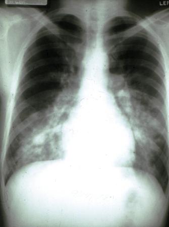
Radiography
The onset of radiographic changes in HAPE can be highly variable.[55] However, most cases eventually develop asymmetric areas of "cotton wool" infiltrates in the mid and lower zones of the lung fields.[56]
Changes tend to begin in the right mid zone and eventually spread across to the left. The apices and costophrenic angles are usually spared. Although prominent pulmonary vasculature may be present, this is a common finding in anyone ascending to altitude.[58] Signs of cardiogenic pulmonary edema are usually absent. Complete resolution of pulmonary infiltrates occurs quickly following recovery.[59][Figure caption and citation for the preceding image starts]: CXR of high-altitude pulmonary edemaPublished with the kind permission of the Wilderness Medical Society [Citation ends].
ECG
Typically shows a sinus tachycardia and changes compatible with acute pulmonary hypertension. These include: right axis deviation and bundle branch block; peaked P waves in leads II, III, and aVF; and an increase in the depth of precordial S waves.[55]
Chest ultrasound and echocardiography
In HAPE, the presence of edema can result in the formation of "comet-tail artifacts" that are visible on ultrasound scanning. These, when assessed over 28 separate lung fields, can provide an objective assessment of pulmonary edema, and can be used to monitor the course of the disease.[57][60] The presence of a patent foramen ovale (present in 25% of the general population) may be associated with an increasing susceptibility to HAPE.[61]
Laboratory investigations
The majority of laboratory investigations are normal in HAPE.
In some cases a small rise in the white blood count occurs; this is due to a mild neutrophil leukocytosis.[55]
HACE
Patients with HACE may be admitted to the hospital, but again this may well not happen due to complexities of rapid evacuation. If admitted with residual symptoms after descent, they should undergo the following tests:
CT brain
In keeping with an increase in intracranial pressure, CT scanning can reveal compression of the ventricles and changes to the gyri and sulci present on the surface of the cerebral hemispheres.
MRI brain
Performed in addition to CT brain
Shows formation of edema in the white matter. This is often concentrated in the splenium of the corpus callosum. Gray matter is largely unaffected by HACE. Changes seen on CT and MRI may take weeks or even months to resolve after clinical recovery.[14][23]
Lumbar puncture
Performed following imaging
An increase in intracranial pressure is often seen in HACE; in advanced cases of HACE, this can exceed normal values by up to 300 mm H2O[62]
Cerebrospinal fluid (CSF) analysis may reveal the presence of red blood cells in severe cases, but in the vast majority of HACE patients CSF will be normal.[4]
Use of this content is subject to our disclaimer