Images and videos
Images


Amebiasis
Life-cycle of Entamoeba histolytica
Reproduced from New England Journal of Medicine (2003); used with permission
See this image in context in the following section/s:

Amebiasis
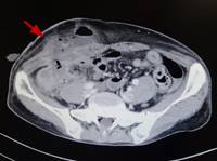
Amebic appendicitis with skin fistula two weeks after appendectomy (enhanced computed tomography).
Original photo from National Center for Global Health and Medicine, Tokyo, Japan.
See this image in context in the following section/s:

Amebiasis
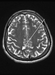
Entamoeba histolytica brain abscess
Reproduced from Transactions of the Royal Society of Tropical Medicine and Hygiene (2007); used with permission
See this image in context in the following section/s:

Amebiasis
Cyst of Entamoeba histolytica: iodine stain of stool sample
Reproduced from Current Concepts (2003); used with permission
See this image in context in the following section/s:
Amebiasis
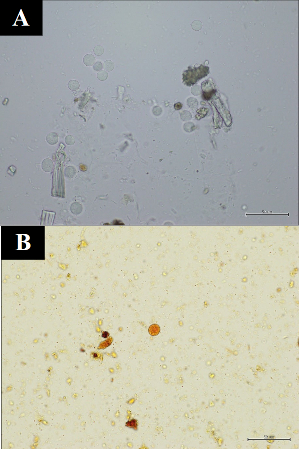
Cyst of Entamoeba histolytica: unstained (A), and iodine stained (B) after formalin-ether concentration of stool sample.
Original photos from National Center for Global Health and Medicine, Tokyo, Japan.
See this image in context in the following section/s:
Amebiasis
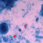
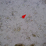
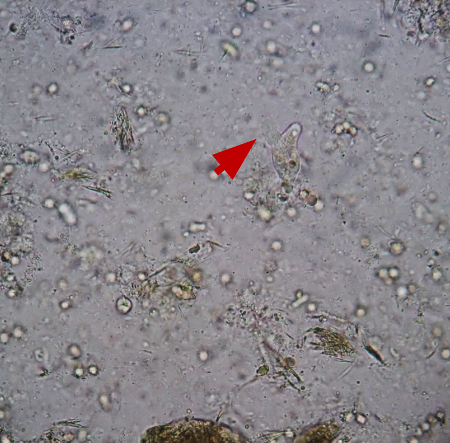
Trophozoite of Entamoeba histolytica with pseudopod (red arrow): direct unstained stool sample.
Original photo from National Center for Global Health and Medicine, Tokyo, Japan.
See this image in context in the following section/s:
Amebiasis
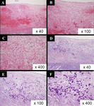
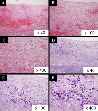
Hematoxilin-Eosin stain (A-C) and Periodic acid-Schiff stain (D-F) of resected appendix of amebic appendicitis. Entamoebas are deeply dyed by Periodic acid-Schiff stain.
Original photo from National Center for Global Health and Medicine, Tokyo, Japan.
See this image in context in the following section/s:

Amebiasis
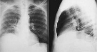
Posterior-anterior and lateral CXR of a patient with amebic liver abscess: CXR findings include elevated right hemidiaphragm and evidence of atelectasis
Reproduced from New England Journal of Medicine (2003); used with permission
See this image in context in the following section/s:

Amebiasis
Trophozoites of Entamoeba histolytica: trichrome stain of stool sample
Reproduced from Clinical Infectious Diseases (1999); used with permission
See this image in context in the following section/s:
Amebiasis
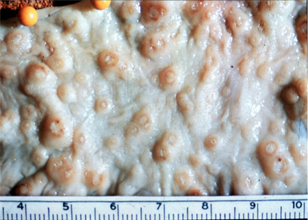
Amebic ulcerations of the colon: colonic ulcers averaging 1 mm to 2 mm in diameter on gross pathology
Reproduced from New England Journal of Medicine (2003); used with permission
See this image in context in the following section/s:
Use of this content is subject to our disclaimer