Images and videos
Images


Leishmaniasis
Female Phlebotomus papatasi sand fly
With kind permission from EdRowtonPhotography.com
See this image in context in the following section/s:
Leishmaniasis
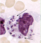
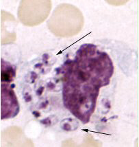
Skin touch preparation showing Leishmania tropica amastigotes. Intact macrophage is practically filled with amastigotes, several of which have a clearly visible nucleus and kinetoplast (arrows)
Image courtesy of CDC; NCID; DPDx
See this image in context in the following section/s:
Leishmaniasis
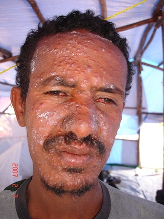
Nodular post-kala-azar dermal leishmaniasis in an Ethiopian patient
Image courtesy of the World Health Organization
See this image in context in the following section/s:
Leishmaniasis
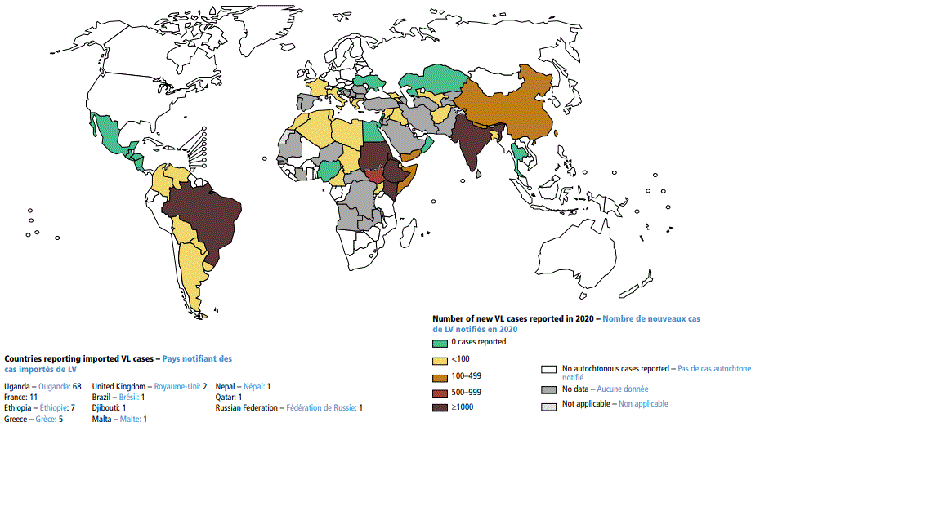
Status of endemicity of visceral leishmaniasis (VL) worldwide, 2020
Global leishmaniasis surveillance: 2019–2020, a baseline for the 2030 roadmap: World Health Organization; 2021. Licence: CC BY-NC-SA 3.0 IGO (https://creativecommons.org/licenses/by-nc-sa/3.0/igo/)
See this image in context in the following section/s:

Leishmaniasis
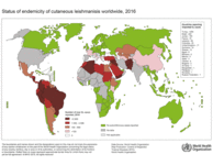
Status of the endemicity of cutaneous leishmaniasis worldwide, 2016
Image courtesy of the World Health Organization
See this image in context in the following section/s:

Leishmaniasis
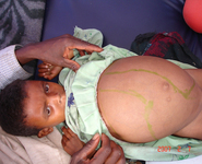
Hepatosplenomegaly in an Ethiopian patient with visceral leishmaniasis
Image courtesy of the World Health Organization
See this image in context in the following section/s:
Leishmaniasis
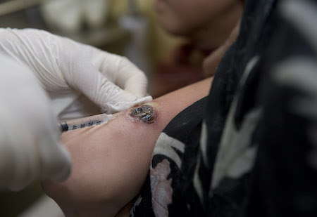
Intralesional injection for the treatment of cutaneous leishmaniasis
Image courtesy of the World Health Organization
See this image in context in the following section/s:
Leishmaniasis
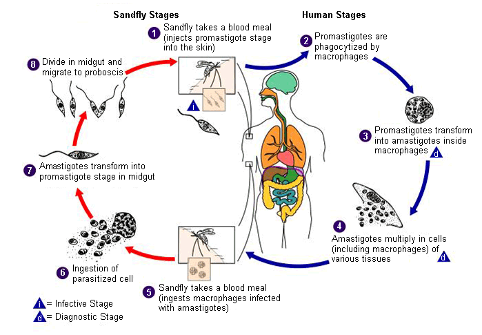
Life cycle of Leishmania species, the causal agents of leishmaniasis
Image courtesy of CDC; A.J. da Silva, PhD; M. Moser
See this image in context in the following section/s:

Leishmaniasis
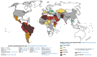
Status of endemicity of cutaneous leishmaniasis (CL) worldwide, 2020
Global leishmaniasis surveillance: 2019–2020, a baseline for the 2030 roadmap: World Health Organization; 2021. Licence: CC BY-NC-SA 3.0 IGO (https://creativecommons.org/licenses/by-nc-sa/3.0/igo/)
See this image in context in the following section/s:

Leishmaniasis
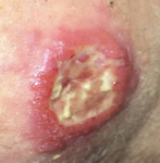
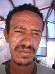
Ulcerative Leishmania braziliensis lesion from a student who traveled to Peru
From the collection of Dr N. Aronson; used with permission
See this image in context in the following section/s:

Leishmaniasis
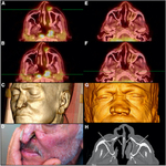
Mucosal leishmaniasis in 2 Brazilian patients. A-D: patient 1 with positron emission tomography/computed tomography (PET/CT) images showing enhancement and subcutaneous thickening adjacent to erosion of the left nasal wing and obliteration of the posterolateral recess (A and B), 3D volume-rendered image of multislice CT data (3D CT) and picture with erosion of the left nasal wing (C and D). F-H: patient 2 with PET/CT images showing preserved glycolytic metabolism of facial structures (E and F), 3D CT with collapse of the nasal pyramids (G), and bone window CT with diffuse thickening of nasal wings and collapse of the nasal pyramid (H)
Am J Trop Med Hyg; CC BY-4.0 (https://creativecommons.org/licenses/by/4.0/)
See this image in context in the following section/s:

Leishmaniasis
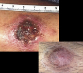
Ulcerative Leishmania mexicana lesion, pre- and post-treatment
From the collection of Dr N. Aronson; used with permission
See this image in context in the following section/s:
Use of this content is subject to our disclaimer