Images and videos
Images


Aortic dissection
Transesophageal echocardiography (transverse aortic section) showing a circumferential dissection of the ascending aorta in a 30-year-old patient with features of Marfan syndrome
Bouzas-Mosquera A, Solla-Buceta M, Fojón-Polanco S. Circumferential aortic dissection. BMJ Case Reports 2009; doi:10.1136/bcr.2007.049908
See this image in context in the following section/s:

Aortic dissection
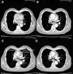
CT of a 71-year-old man showing type II dissecting aneurysm of the ascending aorta. Hematoma around the proximal segment of the ascending aorta (panels A-D) compressed the right pulmonary artery, almost occluding its patency and limiting the perfusion of the reciprocal lung
Stougiannos PN, Mytas DZ, Pyrgakis VN. The changing faces of aortic dissection: an unusual presentation mimicking pulmonary embolism. BMJ Case Reports 2009; doi:10.1136/bcr.2006.104414
See this image in context in the following section/s:
Aortic dissection
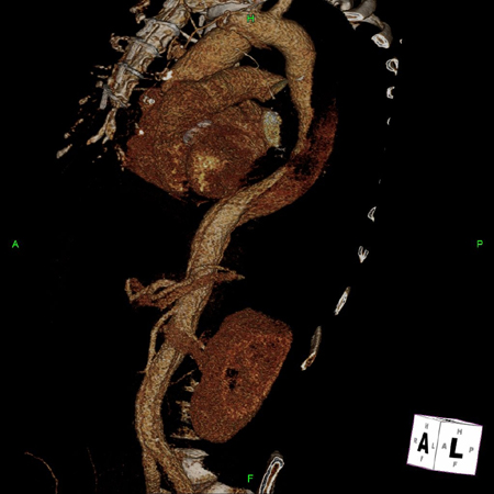
3D CT, distal dissection
From the collection of Dr Eric E. Roselli; used with permission
See this image in context in the following section/s:
Aortic dissection
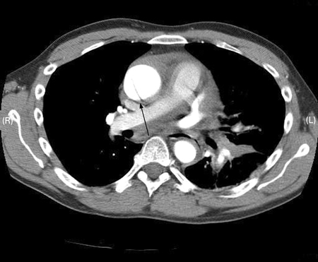
CT scan showing dissecting aneurysm in a 45-year-old patient with Marfan syndrome experiencing chest pain
Sanyal K, Sabanathan K. Chest pain in Marfan syndrome. BMJ Case Reports 2009; doi:10.1136/bcr.07.2008.0431
See this image in context in the following section/s:
Aortic dissection
Proximal dissection
From the collection of Dr Eric E. Roselli; used with permission
See this image in context in the following section/s:
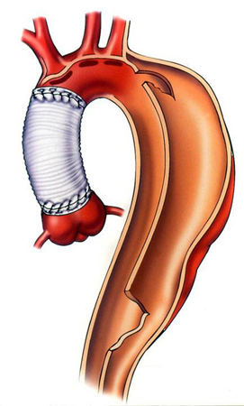
Aortic dissection
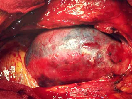
Dissection status post proximal repair with late distal aneurysm
From the collection of Dr Eric E. Roselli; used with permission
See this image in context in the following section/s:
Use of this content is subject to our disclaimer