Images and videos
Images


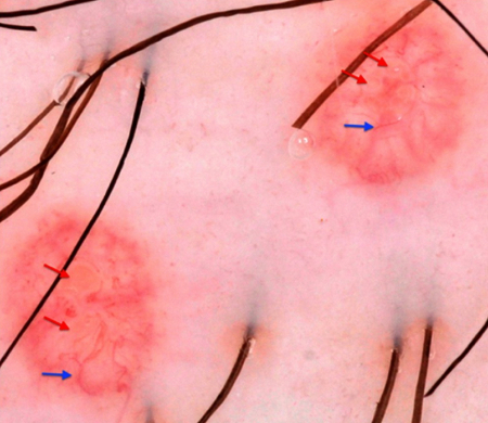
Molluscum contagiosum
Dermatoscopic findings of Molluscum contagiosum. Red arrows indicate white-to-yellow polylobular structures; blue arrows: crown vessels. (Polarized-light dermoscopy, original magnification 10x)
Clinical, Cosmetic and Investigational Dermatology 2019:12 373-381. Originally published by and used with permission from Dove Medical Press Ltd.
See this image in context in the following section/s:
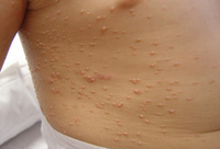
Molluscum contagiosum
Extensive molluscum lesions on the flank of a young child; lesions are flesh to pearly colored with central dells
From the collection of Dr Nanette Silverberg
See this image in context in the following section/s:

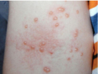
Molluscum contagiosum
Molluscum on the arm of a child with atopic dermatitis demonstrating pearly papules with a central dell and areas of excoriation and erythema due to inflammation
Reproduced with permission from dermnetnz.org
See this image in context in the following section/s:
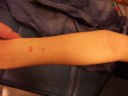
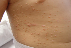
Molluscum contagiosum
Forearm: pearly papules with a central dell and areas of excoriation and erythema due to inflammation
From the collection of Dr Nanette Silverberg
See this image in context in the following section/s:

Molluscum contagiosum
Molluscum contagiosum on the face of an adult, which may be the presenting sign of HIV infection as in this case
Maybury C et al. Peri-oral papules. BMJ. 2013 Feb 11;346:f750; used with permission
See this image in context in the following section/s:
Use of this content is subject to our disclaimer