Differentials
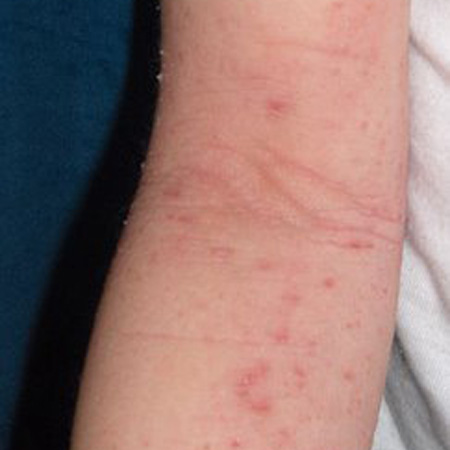
Eczema
SIGNS / SYMPTOMS
Commonly starts in early childhood.[31]
Usually involves the extensor surfaces in infants and flexures in the older child.
May be associated with other atopic diseases such as asthma and allergic rhinitis.[Figure caption and citation for the preceding image starts]: Acute eczema in the antecubital fossa of a 9-year-old girlFrom the personal collection of A. Hebert, MD; used with permission [Citation ends].
INVESTIGATIONS
Histopathology: spongiosis and eosinophils within the dermal inflammatory infiltrate.
Direct immunofluorescence microscopy: absence of granular IgA deposits along the basement membrane zone.
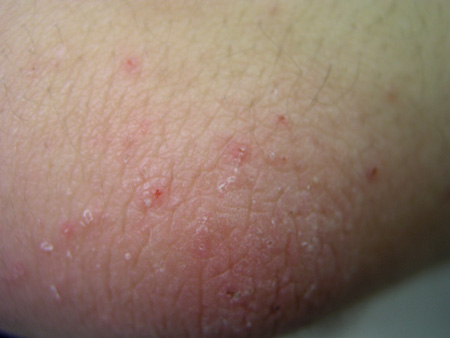
Contact dermatitis
SIGNS / SYMPTOMS
There may be an occupational history of exposure, such as occupations that involve frequent exposure to water.
There may be a history of atopy
Potential triggering factors in allergic contact dermatitis with distribution depending on location of contact with allergen.
[Figure caption and citation for the preceding image starts]: Allergic contact dermatitis to Toxicodendron species From the personal collection of Dr Snehal Desai [Citation ends].

INVESTIGATIONS
Histopathology: spongiosis and eosinophils within the dermal inflammatory infiltrate.
Direct immunofluorescence microscopy: absence of granular IgA deposits along the basement membrane zone.
Scabies
SIGNS / SYMPTOMS
Thin tunnels (burrows) within the epidermis, pruritic papules or nodules, and excoriations mainly located in interdigital areas, armpits, genitals, and nipples.[32]
Pruritus is worse at night.
Highly contagious so family members or close contacts may also be affected.[33]
[Figure caption and citation for the preceding image starts]: Characteristic linear burrows in skinFrom the collection of Laura Ferris, MD, PhD [Citation ends].
INVESTIGATIONS
Microscopic examination: identification of the mite, eggs, or scybala (mite faeces).
Dermoscopic examination: identification of the mite within the burrows.
Prurigo nodularis or subacuta
SIGNS / SYMPTOMS
Patients have pruritic papules, nodules, or excoriations that are not usually grouped.[34]
Mainly found in older people.
INVESTIGATIONS
Direct immunofluorescence microscopy: absence of granular IgA deposits along the basement membrane zone.
Linear IgA bullous dermatosis
SIGNS / SYMPTOMS
Vesicles and blisters in a string of pearls configuration.[35]
Perioral and anogenital regions may be involved.
INVESTIGATIONS
Direct immunofluorescence microscopy: linear IgA deposits along the basement membrane zone.
Indirect immunofluorescence microscopy: linear IgA deposits at the epidermal side using salt split skin as substrate.
Immunoblotting: presence of IgA against LABD97 or LAD-1 antigens.
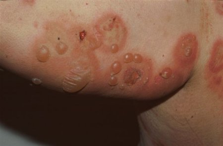
Bullous pemphigoid
SIGNS / SYMPTOMS
Blisters rather than vesicles.
Armpits, upper thighs, and lower abdomen are more commonly affected.
Mucosal lesions may be present in up to 30% of patients.[36]
Older people are most commonly affected.[37]
[Figure caption and citation for the preceding image starts]: Tense, fluid-filled blisters on normal and erythematous skinFrom the collection of Dr Vesna Petronic-Rosic [Citation ends].
INVESTIGATIONS
Direct immunofluorescence microscopy: linear IgG and/or C3 deposits along the basement membrane zone.[38]
Indirect immunofluorescence microscopy: linear IgG deposits at the epidermal side using salt split skin as substrate.
Enzyme-linked immunosorbent assay: IgG against BP180 and/or BP230.
Pemphigus herpetiformis
SIGNS / SYMPTOMS
Arcuate, urticarial, and circinate plaques with peripheral vesicles.
INVESTIGATIONS
Direct immunofluorescence microscopy: intercellular IgG and/or C3 deposits within the epidermis.[39]
Indirect immunofluorescence microscopy: intercellular IgG deposits using monkey oesophagus as substrate.
Enzyme-linked immunosorbent assay: IgG against desmoglein 1.
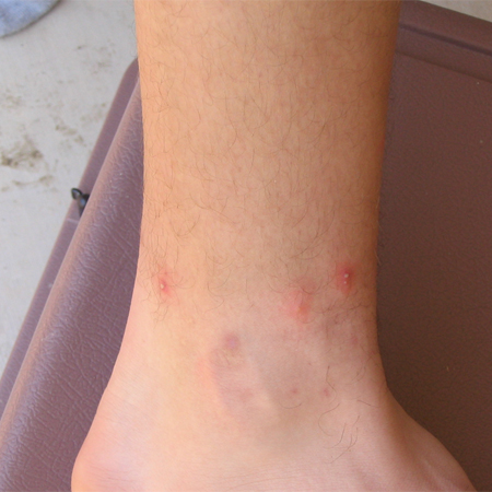
Arthropod bite reactions
SIGNS / SYMPTOMS
Discrete urticarial lesions usually centred by a vesicle on the exposed areas.
Usually self-limiting without chronic course.
History of exposure to arthropod bites.
Seasonality (mostly occurring in spring-summer).
[Figure caption and citation for the preceding image starts]: Pseudopustule formation following fire ant stingCourtesy of Theodore Freeman [Citation ends].
INVESTIGATIONS
Skin biopsy: epidermal spongiosis, focal parakeratosis, and papillary dermal oedema, subepidermal blister with eosinophils, superficial and deep perivascular infiltrate of lymphocytes and eosinophils in dermis.
Direct immunofluorescence microscopy: absence of granular IgA deposits along the basement membrane zone.
Use of this content is subject to our disclaimer