Consider the diagnosis of dermatitis herpetiformis in any patient with a history of a chronic, recurrent, symmetric, polymorphic, and highly pruritic vesicular skin eruption distributed in typical areas, such as the elbows, knees, shoulders, scalp, and buttocks.[2]Görög A, Antiga E, Caproni M, et al. S2k guidelines (consensus statement) for diagnosis and therapy of dermatitis herpetiformis initiated by the European Academy of Dermatology and Venereology (EADV). J Eur Acad Dermatol Venereol. 2021 Jun;35(6):1251-77.
https://onlinelibrary.wiley.com/doi/10.1111/jdv.17183
http://www.ncbi.nlm.nih.gov/pubmed/34004067?tool=bestpractice.com
[3]Ludvigsson JF, Bai JC, Biagi F, et al. Diagnosis and management of adult coeliac disease: guidelines from the British Society of Gastroenterology. Gut. 2014 Aug;63(8):1210-28.
https://gut.bmj.com/content/63/8/1210.long
http://www.ncbi.nlm.nih.gov/pubmed/24917550?tool=bestpractice.com
[11]Caproni M, Antiga E, Melani L, et al. Guidelines for the diagnosis and treatment of dermatitis herpetiformis. J Eur Acad Dermatol Venereol. 2009 Jun;23(6):633-8.
https://onlinelibrary.wiley.com/doi/10.1111/j.1468-3083.2009.03188.x
http://www.ncbi.nlm.nih.gov/pubmed/19470076?tool=bestpractice.com
[16]Hill ID, Fasano A, Guandalini S, et al. NASPGHAN clinical report on the diagnosis and treatment of gluten-related disorders. J Pediatr Gastroenterol Nutr. 2016 Jul;63(1):156-65.
http://www.ncbi.nlm.nih.gov/pubmed/27035374?tool=bestpractice.com
Due to the extreme pruritus, the primary vesicular lesions may be masked by less specific manifestations, such as excoriations, erosions, and crusts.[2]Görög A, Antiga E, Caproni M, et al. S2k guidelines (consensus statement) for diagnosis and therapy of dermatitis herpetiformis initiated by the European Academy of Dermatology and Venereology (EADV). J Eur Acad Dermatol Venereol. 2021 Jun;35(6):1251-77.
https://onlinelibrary.wiley.com/doi/10.1111/jdv.17183
http://www.ncbi.nlm.nih.gov/pubmed/34004067?tool=bestpractice.com
The most common differential diagnoses to rule out include:[2]Görög A, Antiga E, Caproni M, et al. S2k guidelines (consensus statement) for diagnosis and therapy of dermatitis herpetiformis initiated by the European Academy of Dermatology and Venereology (EADV). J Eur Acad Dermatol Venereol. 2021 Jun;35(6):1251-77.
https://onlinelibrary.wiley.com/doi/10.1111/jdv.17183
http://www.ncbi.nlm.nih.gov/pubmed/34004067?tool=bestpractice.com
Atopic dermatitis/eczema
Contact/irritant dermatitis
Multiple folliculitis
Prurigo nodularis/subacuta
Scabies
Arthropod bite reactions
Autoimmune bullous disease (e.g., linear immunoglobulin A [IgA] bullous dermatosis, bullous pemphigoid, pemphigus herpetiformis).
Dermatitis herpetiformis is confirmed by detection of granular IgA deposits at the tips of the dermal papillae or along the basement membrane zone with direct immunofluorescence microscopy.[2]Görög A, Antiga E, Caproni M, et al. S2k guidelines (consensus statement) for diagnosis and therapy of dermatitis herpetiformis initiated by the European Academy of Dermatology and Venereology (EADV). J Eur Acad Dermatol Venereol. 2021 Jun;35(6):1251-77.
https://onlinelibrary.wiley.com/doi/10.1111/jdv.17183
http://www.ncbi.nlm.nih.gov/pubmed/34004067?tool=bestpractice.com
[3]Ludvigsson JF, Bai JC, Biagi F, et al. Diagnosis and management of adult coeliac disease: guidelines from the British Society of Gastroenterology. Gut. 2014 Aug;63(8):1210-28.
https://gut.bmj.com/content/63/8/1210.long
http://www.ncbi.nlm.nih.gov/pubmed/24917550?tool=bestpractice.com
[11]Caproni M, Antiga E, Melani L, et al. Guidelines for the diagnosis and treatment of dermatitis herpetiformis. J Eur Acad Dermatol Venereol. 2009 Jun;23(6):633-8.
https://onlinelibrary.wiley.com/doi/10.1111/j.1468-3083.2009.03188.x
http://www.ncbi.nlm.nih.gov/pubmed/19470076?tool=bestpractice.com
[16]Hill ID, Fasano A, Guandalini S, et al. NASPGHAN clinical report on the diagnosis and treatment of gluten-related disorders. J Pediatr Gastroenterol Nutr. 2016 Jul;63(1):156-65.
http://www.ncbi.nlm.nih.gov/pubmed/27035374?tool=bestpractice.com
History
Take a detailed clinical history from the patient, including questioning regarding:[2]Görög A, Antiga E, Caproni M, et al. S2k guidelines (consensus statement) for diagnosis and therapy of dermatitis herpetiformis initiated by the European Academy of Dermatology and Venereology (EADV). J Eur Acad Dermatol Venereol. 2021 Jun;35(6):1251-77.
https://onlinelibrary.wiley.com/doi/10.1111/jdv.17183
http://www.ncbi.nlm.nih.gov/pubmed/34004067?tool=bestpractice.com
Description of skin symptoms, including severity, location, timing, and duration, as well as how lesions have evolved over time
Involvement of the gastrointestinal system, such as existence of current gastrointestinal symptoms and gastrointestinal medical history (chronic or relapsing abdominal pain, nausea, diarrhoea, constipation, weight loss)
General medical history, including a history of autoimmune or immune-mediated associated diseases (e.g., pernicious anaemia, Hashimoto's thyroiditis, alopecia areata, Addison's disease, or type 1 diabetes mellitus)
Relevant family history, such as coeliac disease, dermatitis herpetiformis, or autoimmune conditions.
Dermatitis herpetiformis usually presents with pruritic lesions that are often preceded by a burning sensation.[2]Görög A, Antiga E, Caproni M, et al. S2k guidelines (consensus statement) for diagnosis and therapy of dermatitis herpetiformis initiated by the European Academy of Dermatology and Venereology (EADV). J Eur Acad Dermatol Venereol. 2021 Jun;35(6):1251-77.
https://onlinelibrary.wiley.com/doi/10.1111/jdv.17183
http://www.ncbi.nlm.nih.gov/pubmed/34004067?tool=bestpractice.com
[3]Ludvigsson JF, Bai JC, Biagi F, et al. Diagnosis and management of adult coeliac disease: guidelines from the British Society of Gastroenterology. Gut. 2014 Aug;63(8):1210-28.
https://gut.bmj.com/content/63/8/1210.long
http://www.ncbi.nlm.nih.gov/pubmed/24917550?tool=bestpractice.com
[11]Caproni M, Antiga E, Melani L, et al. Guidelines for the diagnosis and treatment of dermatitis herpetiformis. J Eur Acad Dermatol Venereol. 2009 Jun;23(6):633-8.
https://onlinelibrary.wiley.com/doi/10.1111/j.1468-3083.2009.03188.x
http://www.ncbi.nlm.nih.gov/pubmed/19470076?tool=bestpractice.com
[17]Antiga E, Caproni M. The diagnosis and treatment of dermatitis herpetiformis. Clin Cosmet Investig Dermatol. 2015 May 13:8:257-65.
https://www.dovepress.com/the-diagnosis-and-treatment-of-dermatitis-herpetiformis-peer-reviewed-fulltext-article-CCID
http://www.ncbi.nlm.nih.gov/pubmed/25999753?tool=bestpractice.com
Pruritus is a universal feature in all patients, which often deeply affects the patient's quality of life.
Skin lesions typically appear as grouped vesicles with a symmetric distribution, most often on extensor surfaces, such as elbows and knees, as well as the shoulders, scalp, back, and buttocks, most often in areas that are frequently exposed to mechanical forces.[2]Görög A, Antiga E, Caproni M, et al. S2k guidelines (consensus statement) for diagnosis and therapy of dermatitis herpetiformis initiated by the European Academy of Dermatology and Venereology (EADV). J Eur Acad Dermatol Venereol. 2021 Jun;35(6):1251-77.
https://onlinelibrary.wiley.com/doi/10.1111/jdv.17183
http://www.ncbi.nlm.nih.gov/pubmed/34004067?tool=bestpractice.com
Less commonly, the posterior nuchal area and/or the hairline may be affected.[2]Görög A, Antiga E, Caproni M, et al. S2k guidelines (consensus statement) for diagnosis and therapy of dermatitis herpetiformis initiated by the European Academy of Dermatology and Venereology (EADV). J Eur Acad Dermatol Venereol. 2021 Jun;35(6):1251-77.
https://onlinelibrary.wiley.com/doi/10.1111/jdv.17183
http://www.ncbi.nlm.nih.gov/pubmed/34004067?tool=bestpractice.com
Patients may also describe involvement of the mouth, such as a history of oral ulcers.[18]Lähteenoja H, Irjala K, Viander M, et al. Oral mucosa is frequently affected in patients with dermatitis herpetiformis. Arch Dermatol. 1998 Jun;134(6):756-8.
http://www.ncbi.nlm.nih.gov/pubmed/9645655?tool=bestpractice.com
Occasionally, dermatitis herpetiformis may occur with atypical cutaneous presentations, including palmar or plantar petechiae, or other rarer signs such as hyperkeratotic lesions of palms and soles, leukocytoclastic vasculitis-like lesions, and prurigo-like lesions.[19]Bonciolini V, Bonciani D, Verdelli A, et al. Newly described clinical and immunopathological feature of dermatitis herpetiformis. Clin Dev Immunol. 2012;2012:967974.
https://www.hindawi.com/journals/jir/2012/967974
http://www.ncbi.nlm.nih.gov/pubmed/22701503?tool=bestpractice.com
Skin lesions usually heal without scarring, but can leave post-inflammatory hyperpigmentation. The disease has a chronic recurrent course, with exacerbations that may be followed by partial remissions of the lesions.
As dermatitis herpetiformis is a specific manifestation of coeliac disease, the condition does not typically present in people who are already on a gluten-free diet for coeliac disease. Around 15% to 20% of people with dermatitis herpetiformis show gastrointestinal symptoms resembling those of coeliac disease, such as diarrhoea, constipation, abdominal bloating or pain, and weight loss. However, most people with dermatitis herpetiformis have only mild gastrointestinal involvement or are asymptomatic.[2]Görög A, Antiga E, Caproni M, et al. S2k guidelines (consensus statement) for diagnosis and therapy of dermatitis herpetiformis initiated by the European Academy of Dermatology and Venereology (EADV). J Eur Acad Dermatol Venereol. 2021 Jun;35(6):1251-77.
https://onlinelibrary.wiley.com/doi/10.1111/jdv.17183
http://www.ncbi.nlm.nih.gov/pubmed/34004067?tool=bestpractice.com
[11]Caproni M, Antiga E, Melani L, et al. Guidelines for the diagnosis and treatment of dermatitis herpetiformis. J Eur Acad Dermatol Venereol. 2009 Jun;23(6):633-8.
https://onlinelibrary.wiley.com/doi/10.1111/j.1468-3083.2009.03188.x
http://www.ncbi.nlm.nih.gov/pubmed/19470076?tool=bestpractice.com
See Coeliac disease.
Children may also present with malabsorption, iron deficiency, and reduced growth rates.[2]Görög A, Antiga E, Caproni M, et al. S2k guidelines (consensus statement) for diagnosis and therapy of dermatitis herpetiformis initiated by the European Academy of Dermatology and Venereology (EADV). J Eur Acad Dermatol Venereol. 2021 Jun;35(6):1251-77.
https://onlinelibrary.wiley.com/doi/10.1111/jdv.17183
http://www.ncbi.nlm.nih.gov/pubmed/34004067?tool=bestpractice.com
Physical examination
Physical examination of patients with dermatitis herpetiformis reveals the presence of symmetric, grouped, polymorphic skin lesions including:[2]Görög A, Antiga E, Caproni M, et al. S2k guidelines (consensus statement) for diagnosis and therapy of dermatitis herpetiformis initiated by the European Academy of Dermatology and Venereology (EADV). J Eur Acad Dermatol Venereol. 2021 Jun;35(6):1251-77.
https://onlinelibrary.wiley.com/doi/10.1111/jdv.17183
http://www.ncbi.nlm.nih.gov/pubmed/34004067?tool=bestpractice.com
[11]Caproni M, Antiga E, Melani L, et al. Guidelines for the diagnosis and treatment of dermatitis herpetiformis. J Eur Acad Dermatol Venereol. 2009 Jun;23(6):633-8.
https://onlinelibrary.wiley.com/doi/10.1111/j.1468-3083.2009.03188.x
http://www.ncbi.nlm.nih.gov/pubmed/19470076?tool=bestpractice.com
[20]Vale ECSD, Dimatos OC, Porro AM, et al. Consensus on the treatment of autoimmune bullous dermatoses: dermatitis herpetiformis and linear IgA bullous dermatosis - Brazilian Society of Dermatology. An Bras Dermatol. 2019 Apr;94(2 suppl 1):48-55.
https://www.ncbi.nlm.nih.gov/pmc/articles/PMC6544034
http://www.ncbi.nlm.nih.gov/pubmed/31166403?tool=bestpractice.com
Erythema
Vesicles
Blisters
Crusts
Urticarial plaques
Papules
Excoriation.
Lesions are usually distributed on typical areas such as the extensor surfaces of upper and lower limbs (mainly elbows [90% of patients] and knees [30% of patients]), buttocks, shoulders, and scalp.[2]Görög A, Antiga E, Caproni M, et al. S2k guidelines (consensus statement) for diagnosis and therapy of dermatitis herpetiformis initiated by the European Academy of Dermatology and Venereology (EADV). J Eur Acad Dermatol Venereol. 2021 Jun;35(6):1251-77.
https://onlinelibrary.wiley.com/doi/10.1111/jdv.17183
http://www.ncbi.nlm.nih.gov/pubmed/34004067?tool=bestpractice.com
[8]Bolotin D, Petronic-Rosic V. Dermatitis herpetiformis. Part I. Epidemiology, pathogenesis, and clinical presentation. J Am Acad Dermatol. 2011 Jun;64(6):1017-24; quiz 1025-6.
http://www.ncbi.nlm.nih.gov/pubmed/21571167?tool=bestpractice.com
[11]Caproni M, Antiga E, Melani L, et al. Guidelines for the diagnosis and treatment of dermatitis herpetiformis. J Eur Acad Dermatol Venereol. 2009 Jun;23(6):633-8.
https://onlinelibrary.wiley.com/doi/10.1111/j.1468-3083.2009.03188.x
http://www.ncbi.nlm.nih.gov/pubmed/19470076?tool=bestpractice.com
[20]Vale ECSD, Dimatos OC, Porro AM, et al. Consensus on the treatment of autoimmune bullous dermatoses: dermatitis herpetiformis and linear IgA bullous dermatosis - Brazilian Society of Dermatology. An Bras Dermatol. 2019 Apr;94(2 suppl 1):48-55.
https://www.ncbi.nlm.nih.gov/pmc/articles/PMC6544034
http://www.ncbi.nlm.nih.gov/pubmed/31166403?tool=bestpractice.com
Less commonly, the patient may have oral mucosa involvement (oral aphthous ulcers), or lesions affecting the posterior nuchal area, and/or the hairline.[2]Görög A, Antiga E, Caproni M, et al. S2k guidelines (consensus statement) for diagnosis and therapy of dermatitis herpetiformis initiated by the European Academy of Dermatology and Venereology (EADV). J Eur Acad Dermatol Venereol. 2021 Jun;35(6):1251-77.
https://onlinelibrary.wiley.com/doi/10.1111/jdv.17183
http://www.ncbi.nlm.nih.gov/pubmed/34004067?tool=bestpractice.com
[18]Lähteenoja H, Irjala K, Viander M, et al. Oral mucosa is frequently affected in patients with dermatitis herpetiformis. Arch Dermatol. 1998 Jun;134(6):756-8.
http://www.ncbi.nlm.nih.gov/pubmed/9645655?tool=bestpractice.com
[20]Vale ECSD, Dimatos OC, Porro AM, et al. Consensus on the treatment of autoimmune bullous dermatoses: dermatitis herpetiformis and linear IgA bullous dermatosis - Brazilian Society of Dermatology. An Bras Dermatol. 2019 Apr;94(2 suppl 1):48-55.
https://www.ncbi.nlm.nih.gov/pmc/articles/PMC6544034
http://www.ncbi.nlm.nih.gov/pubmed/31166403?tool=bestpractice.com
The primary vesicular lesions may in some cases be masked by less specific manifestations, such as excoriations, erosions, and crusts, due to the extensively pruritic nature of the condition.[2]Görög A, Antiga E, Caproni M, et al. S2k guidelines (consensus statement) for diagnosis and therapy of dermatitis herpetiformis initiated by the European Academy of Dermatology and Venereology (EADV). J Eur Acad Dermatol Venereol. 2021 Jun;35(6):1251-77.
https://onlinelibrary.wiley.com/doi/10.1111/jdv.17183
http://www.ncbi.nlm.nih.gov/pubmed/34004067?tool=bestpractice.com
[Figure caption and citation for the preceding image starts]: Characteristic pruritic papulovesicular rash of dermatitis herpetiformis, affecting the elbowsCID - ISM/Science Photo Library; used with permission [Citation ends].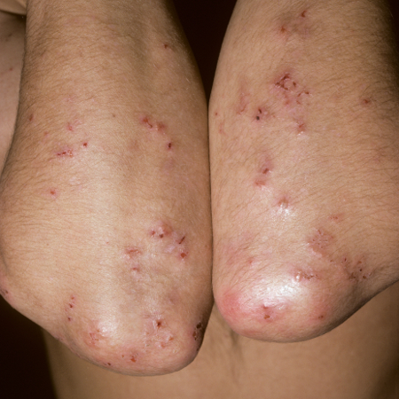 [Figure caption and citation for the preceding image starts]: Characteristic pruritic papulovesicular rash of dermatitis herpetiformis, affecting back and buttocksScience Photo Library; used with permission [Citation ends].
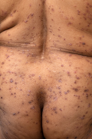 [Figure caption and citation for the preceding image starts]: Aphthous ulcer: a less common manifestation of dermatitis herpetiformisFrom the collection of Dr Michaell A. Huber, UTHSCSA School of Dentistry; used with permission [Citation ends].
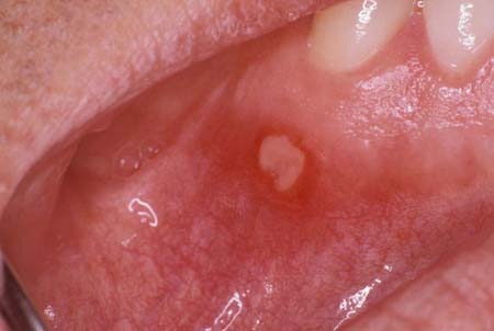
Dermatitis herpetiformis may present atypically, with signs including palmar or plantar petechiae, or rarer signs such as hyperkeratotic lesions of palms and soles, leukocytoclastic vasculitis-like lesions, and prurigo-like lesions.[19]Bonciolini V, Bonciani D, Verdelli A, et al. Newly described clinical and immunopathological feature of dermatitis herpetiformis. Clin Dev Immunol. 2012;2012:967974.
https://www.hindawi.com/journals/jir/2012/967974
http://www.ncbi.nlm.nih.gov/pubmed/22701503?tool=bestpractice.com
Skin lesions usually heal without scarring, but can leave post-inflammatory hyperpigmentation.[2]Görög A, Antiga E, Caproni M, et al. S2k guidelines (consensus statement) for diagnosis and therapy of dermatitis herpetiformis initiated by the European Academy of Dermatology and Venereology (EADV). J Eur Acad Dermatol Venereol. 2021 Jun;35(6):1251-77.
https://onlinelibrary.wiley.com/doi/10.1111/jdv.17183
http://www.ncbi.nlm.nih.gov/pubmed/34004067?tool=bestpractice.com
Around 15% to 20% of patients with dermatitis herpetiformis show gastrointestinal symptoms typical of coeliac disease; these patients may also have signs of malabsorption, which include:[2]Görög A, Antiga E, Caproni M, et al. S2k guidelines (consensus statement) for diagnosis and therapy of dermatitis herpetiformis initiated by the European Academy of Dermatology and Venereology (EADV). J Eur Acad Dermatol Venereol. 2021 Jun;35(6):1251-77.
https://onlinelibrary.wiley.com/doi/10.1111/jdv.17183
http://www.ncbi.nlm.nih.gov/pubmed/34004067?tool=bestpractice.com
Initial investigations
Order the following investigations in a patient with suspected dermatitis herpetiformis:[2]Görög A, Antiga E, Caproni M, et al. S2k guidelines (consensus statement) for diagnosis and therapy of dermatitis herpetiformis initiated by the European Academy of Dermatology and Venereology (EADV). J Eur Acad Dermatol Venereol. 2021 Jun;35(6):1251-77.
https://onlinelibrary.wiley.com/doi/10.1111/jdv.17183
http://www.ncbi.nlm.nih.gov/pubmed/34004067?tool=bestpractice.com
[11]Caproni M, Antiga E, Melani L, et al. Guidelines for the diagnosis and treatment of dermatitis herpetiformis. J Eur Acad Dermatol Venereol. 2009 Jun;23(6):633-8.
https://onlinelibrary.wiley.com/doi/10.1111/j.1468-3083.2009.03188.x
http://www.ncbi.nlm.nih.gov/pubmed/19470076?tool=bestpractice.com
[20]Vale ECSD, Dimatos OC, Porro AM, et al. Consensus on the treatment of autoimmune bullous dermatoses: dermatitis herpetiformis and linear IgA bullous dermatosis - Brazilian Society of Dermatology. An Bras Dermatol. 2019 Apr;94(2 suppl 1):48-55.
https://www.ncbi.nlm.nih.gov/pmc/articles/PMC6544034
http://www.ncbi.nlm.nih.gov/pubmed/31166403?tool=bestpractice.com
[21]Al-Toma A, Volta U, Auricchio R, et al. European Society for the Study of Coeliac Disease (ESsCD) guideline for coeliac disease and other gluten-related disorders. United European Gastroenterol J. 2019 Jun;7(5):583-613.
https://onlinelibrary.wiley.com/doi/10.1177/2050640619844125
http://www.ncbi.nlm.nih.gov/pubmed/31210940?tool=bestpractice.com
Skin biopsy of lesional skin for histopathological examination and of perilesional skin for direct immunofluorescence microscopy.
The optimal biopsy site is perilesional, uninvolved skin because direct immunofluorescence microscopy performed on lesional biopsies rather than perilesional skin often leads to false-negative results.[2]Görög A, Antiga E, Caproni M, et al. S2k guidelines (consensus statement) for diagnosis and therapy of dermatitis herpetiformis initiated by the European Academy of Dermatology and Venereology (EADV). J Eur Acad Dermatol Venereol. 2021 Jun;35(6):1251-77.
https://onlinelibrary.wiley.com/doi/10.1111/jdv.17183
http://www.ncbi.nlm.nih.gov/pubmed/34004067?tool=bestpractice.com
[22]Zone JJ, Meyer LJ, Petersen MJ. Deposition of granular IgA relative to clinical lesions in dermatitis herpetiformis. Arch Dermatol. 1996 Aug;132(8):912-8.
http://www.ncbi.nlm.nih.gov/pubmed/8712841?tool=bestpractice.com
Blood tests to detect immunoglobulin A-tissue transglutaminase (IgA-tTG), and if this is negative, test for the presence of endomysial antibody (EMA).
Direct immunofluorescence microscopy is the definitive investigation for the diagnosis of dermatitis herpetiformis and almost universally shows granular deposition of IgA at the tips of dermal papillae and/or along the dermal-epidermal junction.[2]Görög A, Antiga E, Caproni M, et al. S2k guidelines (consensus statement) for diagnosis and therapy of dermatitis herpetiformis initiated by the European Academy of Dermatology and Venereology (EADV). J Eur Acad Dermatol Venereol. 2021 Jun;35(6):1251-77.
https://onlinelibrary.wiley.com/doi/10.1111/jdv.17183
http://www.ncbi.nlm.nih.gov/pubmed/34004067?tool=bestpractice.com
[11]Caproni M, Antiga E, Melani L, et al. Guidelines for the diagnosis and treatment of dermatitis herpetiformis. J Eur Acad Dermatol Venereol. 2009 Jun;23(6):633-8.
https://onlinelibrary.wiley.com/doi/10.1111/j.1468-3083.2009.03188.x
http://www.ncbi.nlm.nih.gov/pubmed/19470076?tool=bestpractice.com
[16]Hill ID, Fasano A, Guandalini S, et al. NASPGHAN clinical report on the diagnosis and treatment of gluten-related disorders. J Pediatr Gastroenterol Nutr. 2016 Jul;63(1):156-65.
http://www.ncbi.nlm.nih.gov/pubmed/27035374?tool=bestpractice.com
[20]Vale ECSD, Dimatos OC, Porro AM, et al. Consensus on the treatment of autoimmune bullous dermatoses: dermatitis herpetiformis and linear IgA bullous dermatosis - Brazilian Society of Dermatology. An Bras Dermatol. 2019 Apr;94(2 suppl 1):48-55.
https://www.ncbi.nlm.nih.gov/pmc/articles/PMC6544034
http://www.ncbi.nlm.nih.gov/pubmed/31166403?tool=bestpractice.com
If a patient with high suspicion of dermatitis herpetiformis tests negative at direct immunofluorescence microscopy, request a repeat skin biopsy; false-negative results after repeated biopsies are very rare.[23]Beutner EH, Baughman RD, Austin BM, et al. A case of dermatitis herpetiformis with IgA endomysial antibodies but negative direct immunofluorescent findings. J Am Acad Dermatol. 2000 Aug;43(2 pt 2):329-32.
http://www.ncbi.nlm.nih.gov/pubmed/10901714?tool=bestpractice.com
Histopathological examination usually shows accumulation of neutrophils at the dermal papillae and a dermal-epidermal detachment; however, in about one third of patients, histopathological examination is not specific, showing only perivascular infiltration of lymphocytes.[24]Warren SJ, Cockerell CJ. Characterization of a subgroup of patients with dermatitis herpetiformis with nonclassical histologic features. Am J Dermatopathol. 2002 Aug;24(4):305-8.
http://www.ncbi.nlm.nih.gov/pubmed/12142608?tool=bestpractice.com
Use serological testing to aid the diagnosis, but it should not be used alone to diagnose the disease.[2]Görög A, Antiga E, Caproni M, et al. S2k guidelines (consensus statement) for diagnosis and therapy of dermatitis herpetiformis initiated by the European Academy of Dermatology and Venereology (EADV). J Eur Acad Dermatol Venereol. 2021 Jun;35(6):1251-77.
https://onlinelibrary.wiley.com/doi/10.1111/jdv.17183
http://www.ncbi.nlm.nih.gov/pubmed/34004067?tool=bestpractice.com
IgA-tTG detected by enzyme-linked immunosorbent assay (ELISA) is present in up to 95% of patients and should be ordered as the initial serological test.[25]Dahlbom I, Korponay-Szabó IR, Kovács JB, et al. Prediction of clinical and mucosal severity of coeliac disease and dermatitis herpetiformis by quantification of IgA/IgG serum antibodies to tissue transglutaminase. J Pediatr Gastroenterol Nutr. 2010 Feb;50(2):140-6.
http://www.ncbi.nlm.nih.gov/pubmed/19841593?tool=bestpractice.com
Order EMA, detected by indirect immunofluorescence, if IgA-tTG antibody testing is negative.
EMA testing has a sensitivity ranging from 60% to 90%.[26]Norsa L, Shamir R, Zevit N, et al. Cardiovascular disease risk factor profiles in children with celiac disease on gluten-free diets. World J Gastroenterol. 2013 Sep 14;19(34):5658-64.
https://www.wjgnet.com/1007-9327/full/v19/i34/5658.htm
http://www.ncbi.nlm.nih.gov/pubmed/24039358?tool=bestpractice.com
[27]Kumar V, Jarzabek-Chorzelska M, Sulej J, et al. Tissue transglutaminase and endomysial antibodies-diagnostic markers of gluten-sensitive enteropathy in dermatitis herpetiformis. Clin Immunol. 2001 Mar;98(3):378-82.
http://www.ncbi.nlm.nih.gov/pubmed/11237562?tool=bestpractice.com
[28]Volta U, Molinaro N, De Franchis R, et al. Correlation between IgA antiendomysial antibodies and subtotal villous atrophy in dermatitis herpetiformis. J Clin Gastroenterol. 1992 Jun;14(4):298-301.
http://www.ncbi.nlm.nih.gov/pubmed/1607605?tool=bestpractice.com
However, these autoantibodies are specific for the diagnosis of coeliac disease rather than of dermatitis herpetiformis; moreover, up to 10% of patients with dermatitis herpetiformis do not have detectable autoantibodies.[7]Antiga E, Bonciolini V, Cazzaniga S, et al. Female patients with dermatitis herpetiformis show a reduced diagnostic delay and have higher sensitivity rates at autoantibody testing for celiac disease. Biomed Res Int. 2019 Dec 29:2019:6307035.
https://www.hindawi.com/journals/bmri/2019/6307035
http://www.ncbi.nlm.nih.gov/pubmed/32090062?tool=bestpractice.com
Other investigations
Order genetic testing for human leukocyte antigen (HLA)-DQ2 and HLA-DQ8 if dermatitis herpetiformis is not confirmed by initial investigations.[2]Görög A, Antiga E, Caproni M, et al. S2k guidelines (consensus statement) for diagnosis and therapy of dermatitis herpetiformis initiated by the European Academy of Dermatology and Venereology (EADV). J Eur Acad Dermatol Venereol. 2021 Jun;35(6):1251-77.
https://onlinelibrary.wiley.com/doi/10.1111/jdv.17183
http://www.ncbi.nlm.nih.gov/pubmed/34004067?tool=bestpractice.com
[11]Caproni M, Antiga E, Melani L, et al. Guidelines for the diagnosis and treatment of dermatitis herpetiformis. J Eur Acad Dermatol Venereol. 2009 Jun;23(6):633-8.
https://onlinelibrary.wiley.com/doi/10.1111/j.1468-3083.2009.03188.x
http://www.ncbi.nlm.nih.gov/pubmed/19470076?tool=bestpractice.com
[21]Al-Toma A, Volta U, Auricchio R, et al. European Society for the Study of Coeliac Disease (ESsCD) guideline for coeliac disease and other gluten-related disorders. United European Gastroenterol J. 2019 Jun;7(5):583-613.
https://onlinelibrary.wiley.com/doi/10.1177/2050640619844125
http://www.ncbi.nlm.nih.gov/pubmed/31210940?tool=bestpractice.com
This test cannot confirm the diagnosis, but a negative test can be used to rule out the condition in these patients due to its high negative predictive rate.[2]Görög A, Antiga E, Caproni M, et al. S2k guidelines (consensus statement) for diagnosis and therapy of dermatitis herpetiformis initiated by the European Academy of Dermatology and Venereology (EADV). J Eur Acad Dermatol Venereol. 2021 Jun;35(6):1251-77.
https://onlinelibrary.wiley.com/doi/10.1111/jdv.17183
http://www.ncbi.nlm.nih.gov/pubmed/34004067?tool=bestpractice.com
The HLA-DQ2 and HLA-DQ8 haplotypes are present in 95% and 5%, respectively, of people with dermatitis herpetiformis.[10]Zone JJ. Skin manifestations of celiac disease. Gastroenterology. 2005 Apr;128(4 suppl 1):S87-91.
https://www.gastrojournal.org/article/S0016-5085(05)00195-2/fulltext
http://www.ncbi.nlm.nih.gov/pubmed/15825132?tool=bestpractice.com
[11]Caproni M, Antiga E, Melani L, et al. Guidelines for the diagnosis and treatment of dermatitis herpetiformis. J Eur Acad Dermatol Venereol. 2009 Jun;23(6):633-8.
https://onlinelibrary.wiley.com/doi/10.1111/j.1468-3083.2009.03188.x
http://www.ncbi.nlm.nih.gov/pubmed/19470076?tool=bestpractice.com
Perform a full blood count with or without ferritin levels to assess for anaemia in patients with confirmed dermatitis herpetiformis and to obtain baseline levels prior to commencement of treatment.[2]Görög A, Antiga E, Caproni M, et al. S2k guidelines (consensus statement) for diagnosis and therapy of dermatitis herpetiformis initiated by the European Academy of Dermatology and Venereology (EADV). J Eur Acad Dermatol Venereol. 2021 Jun;35(6):1251-77.
https://onlinelibrary.wiley.com/doi/10.1111/jdv.17183
http://www.ncbi.nlm.nih.gov/pubmed/34004067?tool=bestpractice.com
Patients with bowel involvement may have reduced haemoglobin levels and associated microcytic hypochromic red cells.
Consider referral for gastrological consultation once the diagnosis of dermatitis herpetiformis is confirmed to assess gastrointestinal status and for consideration of gastroscopy with duodenal biopsy (unless a diagnosis of coeliac disease has been confirmed).[2]Görög A, Antiga E, Caproni M, et al. S2k guidelines (consensus statement) for diagnosis and therapy of dermatitis herpetiformis initiated by the European Academy of Dermatology and Venereology (EADV). J Eur Acad Dermatol Venereol. 2021 Jun;35(6):1251-77.
https://onlinelibrary.wiley.com/doi/10.1111/jdv.17183
http://www.ncbi.nlm.nih.gov/pubmed/34004067?tool=bestpractice.com
The majority of patients with dermatitis herpetiformis (up to 70%) have a mild enteropathy with mild villous atrophy; the remainder may show severe intestinal involvement or, in few cases, the presence of normal bowel mucosa.[15]Mansikka E, Hervonen K, Kaukinen K, et al. Prognosis of dermatitis herpetiformis patients with and without villous atrophy at diagnosis. Nutrients. 2018 May 19;10(5):641.
https://www.mdpi.com/2072-6643/10/5/641
http://www.ncbi.nlm.nih.gov/pubmed/29783727?tool=bestpractice.com
Also consider assessment for associated conditions such as:[2]Görög A, Antiga E, Caproni M, et al. S2k guidelines (consensus statement) for diagnosis and therapy of dermatitis herpetiformis initiated by the European Academy of Dermatology and Venereology (EADV). J Eur Acad Dermatol Venereol. 2021 Jun;35(6):1251-77.
https://onlinelibrary.wiley.com/doi/10.1111/jdv.17183
http://www.ncbi.nlm.nih.gov/pubmed/34004067?tool=bestpractice.com
Other associated conditions to be aware of include Addison's disease, vitiligo, alopecia areata, and several other autoimmune diseases.[2]Görög A, Antiga E, Caproni M, et al. S2k guidelines (consensus statement) for diagnosis and therapy of dermatitis herpetiformis initiated by the European Academy of Dermatology and Venereology (EADV). J Eur Acad Dermatol Venereol. 2021 Jun;35(6):1251-77.
https://onlinelibrary.wiley.com/doi/10.1111/jdv.17183
http://www.ncbi.nlm.nih.gov/pubmed/34004067?tool=bestpractice.com
[29]Reunala T, Collin P. Diseases associated with dermatitis herpetiformis. Br J Dermatol. 1997 Mar;136(3):315-8.
http://www.ncbi.nlm.nih.gov/pubmed/9115907?tool=bestpractice.com
Pitfalls in diagnosis
Although the diagnosis of dermatitis herpetiformis can easily be confirmed by direct immunofluorescence microscopy, clinical suspicion may not arise in the first instance due to the rarity of the condition as well as the polymorphic clinical appearance. This can lead to delays in diagnosis, with the condition often being overlooked in favour of more common skin diseases.
Moreover, if serological testing for IgA-tTG or EMA is performed before direct immunofluorescence microscopy and results are negative, the diagnosis of dermatitis herpetiformis may be wrongly excluded.
It is also worth noting that some patients with coeliac disease who have skin manifestations other than dermatitis herpetiformis (e.g., psoriasis, eczema, and granuloma annulare) may demonstrate granular IgA deposits in perilesional skin.[30]Bonciolini V, Antiga E, Bianchi B, et al. Granular IgA deposits in the skin of patients with coeliac disease: is it always dermatitis herpetiformis? Acta Derm Venereol. 2019 Jan 1;99(1):78-83.
http://www.ncbi.nlm.nih.gov/pubmed/29972219?tool=bestpractice.com
In these cases, misdiagnosis of dermatitis herpetiformis can occur.
It is important to give only symptomatic therapy until all diagnostic steps have been carried out, as gluten-free diet and dapsone treatment may modify diagnostic results.[2]Görög A, Antiga E, Caproni M, et al. S2k guidelines (consensus statement) for diagnosis and therapy of dermatitis herpetiformis initiated by the European Academy of Dermatology and Venereology (EADV). J Eur Acad Dermatol Venereol. 2021 Jun;35(6):1251-77.
https://onlinelibrary.wiley.com/doi/10.1111/jdv.17183
http://www.ncbi.nlm.nih.gov/pubmed/34004067?tool=bestpractice.com
Gluten-free diet can alter the presence of granular IgA deposits detected by direct immunofluorescence microscopy.
Dapsone may clear clinical lesions, therefore making histology unreliable at confirming the diagnosis.