History and exam
Key diagnostic factors
common
presence of risk factors
A young age at presentation (<2 years) and having a positive family history of XP and/or parental consanguinity are strong risk factors for XP.
photosensitivity
Around 60% of patients have a history of photosensitivity, presenting as skin erythema, burning, and blistering with minimal sun exposure.[3][25] Erythema can persist for weeks before resolving. This is a common presenting feature in patients with XP subtypes XPA, XPB, XPD, XPF, and XPG.[7]
[Figure caption and citation for the preceding image starts]: A child with XP (subtype XPD) at 9 months of age with severe blistering erythema of the malar area following minimal sun exposure. Note the sparing of her forehead and eyes that were protected by a hatBradford PT et al. J Med Genet 2011;48:168-76; used with permission [Citation ends].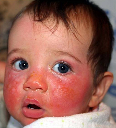
[Figure caption and citation for the preceding image starts]: Relatively protected skin on buttocks (covered by double clothing layers) of 35-year old man shows sparing of the pigmented skin lesions (lentigos) of photodamageRizza ERH et al. J Invest Dermatol 2021 Apr;141(4S):976-84; used with permission [Citation ends].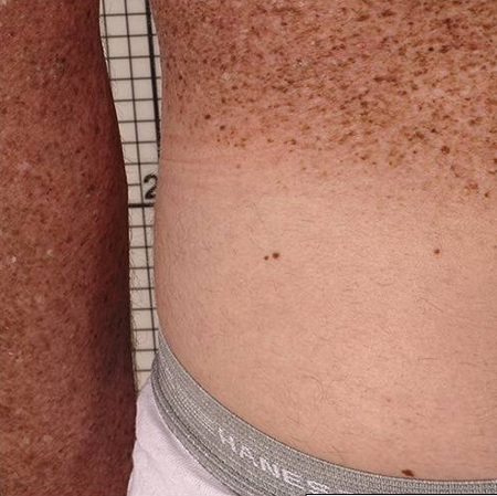
Some 40% of patients do not have acute burning with minimal sun exposure. These patients will tan normally and have many freckle-like lesions (lentigines) on sun-exposed areas.[23]
photophobia
Acute and suggestive of ocular involvement. May be associated with keratitis.
prominent conjunctival injection
May be suggestive of ocular involvement.
excessive freckle-like skin pigmentation (lentigines)
The skin of children with XP shows early and accelerated skin aging.
Pronounced skin pigmentation especially before age 2 years, including freckle-like lesions (lentigines: atypical lesions that are variable intensity of brown in color with irregular shapes, sizes, and borders) over sun-exposed areas. This finding is common in XP (seen in all subtypes) and rare in the general population.[14]
skin xerosis
The skin of children with XP shows early and accelerated skin aging.[7][23]
The skin aging is similar to that seen following lifelong sun exposure in those spending a large amount of time outdoors, such as in farmers and sailors, with xerosis, atrophy, wrinkling, telangiectasia, and dyspigmentation.[1]
skin atrophy
The skin of children with XP shows early and accelerated skin aging.[7][23]This includes the eyelids.
The skin aging is similar to that seen following lifelong sun exposure in those spending a large amount of time outdoors, such as in farmers and sailors, with xerosis, atrophy, wrinkling, telangiectasia, and dyspigmentation.[1]
The manifestations of poikiloderma may be less prominent or less noticeable in people with darker Fitzpatrick skin phototypes.[31]
skin wrinkling
The skin of children with XP shows early and accelerated skin aging.[7][23]
The skin aging is similar to that seen following lifelong sun exposure in those spending a large amount of time outdoors, such as in farmers and sailors, with xerosis, atrophy, wrinkling, telangiectasia, and dyspigmentation.[1]
skin telangiectasia
The skin of children with XP shows early and accelerated skin aging.
The skin aging is similar to that seen following lifelong sun exposure in those spending a large amount of time outdoors, such as in farmers and sailors, with xerosis, atrophy, wrinkling, telangiectasia, and dyspigmentation.[1]
signs of skin cancer
Patients aged <20 years have a more than a 1000-fold increased risk of non-melanoma or melanoma skin cancer.[25] The average age at onset of first skin cancer is <10 years old.[14] Basal cell carcinoma, squamous cell carcinoma, and malignant melanoma within the first decade of life is highly suggestive of XP.[7][25] These can manifest as non-healing wounds, growing papules or nodules on the skin, and/or unusual cutaneous pigmentation.
Patients with subtypes XPC, XPE, and XPV may be less likely to develop severe sunburn reactions, but have large numbers of freckle-like lesions on sun-exposed skin (including lips) and increased skin cancer risk. In particular, XPC subtype shows significant exposed-site lentigines. XPE and XPV subtypes often present in adulthood with early onset of skin cancer.[4][7]
[Figure caption and citation for the preceding image starts]: A child with XP (subtype XPC) at age 2 years. Her parents reported that she did not sunburn easily, but she developed multiple hyperpigmented macules on her face. A rapidly growing squamous cell carcinoma or keratoacanthoma grew on her upper lip and a pre-cancerous lesion appeared on her foreheadBradford PT et al. J Med Genet 2011;48:168-76; used with permission [Citation ends].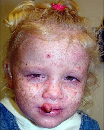
conjunctivitis
A sign of chronic sun-induced damage to the eyes.[27]
corneal neovascularization
A sign of chronic sun-induced damage to the eyes.[27]
dry eye
A sign of chronic sun-induced damage to the eyes.[27]
keratitis/corneal scarring
A sign of chronic severe sun-induced damage to the cornea, which results in photophobia, and painful red, watery eyes. Exposure keratitis leads to corneal opacification or vascularization.[27]
ectropion
A sign of chronic sun-induced damage to the eyes.[27]
blepharitis
A sign of sun-induced damage to the eyes.[27]
conjunctival melanosis
A sign of sun-induced damage to the eyes.[27]
cataract
A sign of sun-induced damage to the eyes.[27]
signs of malignancy over the lids, conjunctiva, or cornea
Associated with sun-induced damage to the eyes.[27]
uncommon
entropion
A sign of chronic sun-induced damage to the eyes.[27]
diminished or absent deep tendon stretch reflex
An early sign of neurologic involvement in XP.
While any XP subtype may be affected, neurodegeneration appears to be most common in subtypes XPA, XPD followed by XPB, XPG, and XPF; and least common in XPC, XPE, and XPV.[7] Adult-onset neurodegeneration has been reported in patients with mutations in the XPF (ERCC4) gene.[30]
sensorineural deafness
May be a feature of neurologic involvement. Where present, this is progressive and irreversible.
While any XP subtype may be affected, neurodegeneration appears to be most common in subtypes XPA, XPD followed by XPB, XPG, and XPF; and least common in XPC, XPE, and XPV.[7] Adult-onset neurodegeneration has been reported in patients with mutations in the XPF (ERCC4) gene.[30]
cognitive impairment
May be a feature of neurologic involvement. Where present, this is progressive and irreversible.
While any XP subtype may be affected, neurodegeneration appears to be most common in subtypes XPA, XPD followed by XPB, XPG, and XPF; and least common in XPC, XPE, and XPV.[7] Adult-onset neurodegeneration has been reported in patients with mutations in the XPF (ERCC4) gene.[30]
ataxia
May be a feature of neurologic involvement.
While any XP subtype may be affected, neurodegeneration appears to be most common in subtypes XPA, XPD followed by XPB, XPG, and XPF; and least common in XPC, XPE, and XPV.[7] Adult-onset neurodegeneration has been reported in patients with mutations in the XPF (ERCC4) gene.[30]
acquired microcephaly
May be a feature of neurologic involvement.
While any XP subtype may be affected, neurodegeneration appears to be most common in subtypes XPA, XPD followed by XPB, XPG, and XPF; and least common in XPC, XPE, and XPV.[7] Adult-onset neurodegeneration has been reported in patients with mutations in the XPF (ERCC4) gene.[30]
other neurodegenerative manifestations
In addition to sensorineural deafness, cognitive impairment, ataxia, and acquired microcephaly, other neurodegenerative manifestations occurring in patients with XP include decreased visual acuity, developmental delay, speech delay, and hyporeflexia.
While any XP subtype may be affected, neurodegeneration appears to be most common in subtypes XPA, XPD followed by XPB, XPG, and XPF; and least common in XPC, XPE, and XPV.[7] Adult-onset neurodegeneration has been reported in patients with mutations in the XPF (ERCC4) gene.[30]
Risk factors
strong
age <2 years
Most patients develop early onset freckling before the age of 2 years.[14] Patients can present with severe and exaggerated sunburn with minimal sun exposure (40%) or with early and excessive freckle-like lesions (lentigines) in sun-exposed areas (100%).[23] Patients who do not have acute burning with minimal sun exposure will tan normally and have many lentigines on sun-exposed areas.[23]
When adults present with XP, they tend to be aged between 20 and 40 years with numerous premalignant or malignant skin cancers.[2]
positive family history
Given that XP is an autosomal recessive disorder with 100% penetrance, there may be a family history of XP in a sibling.[20] However, no family history of XP does not preclude the diagnosis. The risk of having a child with XP is 1 in 4 for each pregnancy when both parents are clinically normal heterozygous carriers.[7]
parental consanguinity
Since XP is inherited in a recessive pattern, there is a higher frequency of this condition in consanguineous populations.
In the absence of a new pathological genetic variant in the proband, each parent of a child with XP will be a normal-appearing carrier for the causative pathological genetic variant. With each additional pregnancy, the risk of having another affected child, an asymptomatic heterozygous carrier, or a genetically unaffected child is 25%, 50%, or 25%, respectively.[7]
weak
Japanese ancestry
Certain geographical regions have a relatively higher incidence of XP. For example, the incidence of XP is estimated to be 1 per million live births in the US, 2.3 per million live births in western Europe, 17.5 per million live births in the Middle East, and 45 per million live births in Japan.[5][6][7] Studies have shown that the incidence of XP is increased in countries where consanguinity is common such as Tunisia, Morocco, Libya, and Pakistan.[6][8][9][10][11]
Subtype XPC is the most common subtype in the US, Europe, and Africa.[12]
Subtype XPA is the most common subtype in China and Japan.[2][12] More than 50% of patients with XP in Japan have the XPA subtype.[13]
Use of this content is subject to our disclaimer