Tests
1st tests to order
posteroanterior and lateral chest x-ray
Test
Indicated in any patient with dyspnea. A posteroanterior chest x-ray and lateral view (now less commonly done) is the first test for this condition. It may confirm the clinical suspicion of, or incidentally reveal, a pleural effusion, but should usually prompt pleural ultrasound.[12][17]
An effusion as small as 50 mL can be seen on the lateral film and more than a few hundred milliliters will be visible on the posteroanterior film.[2]
May show an effusion before any other symptoms arise.[Figure caption and citation for the preceding image starts]: Left-sided pleural effusionFrom the collection of R Light [Citation ends].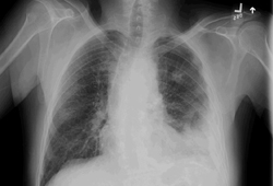
Result
blunting of the costophrenic angles
pleural ultrasound
Test
Useful in locating an area of fluid collection for thoracentesis, especially if the effusion is loculated or small.
Can detect septations within a pleural collection.
Can provide information regarding the nature of the effusion, such as echogenic swirling in exudates.
More sensitive and specific than chest x-ray for pleural effusion detection. It is commonly used as a point of care test to guide intervention with strong evidence suggesting this improves safety and can guide management decisions.[20][24][25][26][17]
Can identify 5 to 10 mL of fluid.[61]
Result
fluid in the pleural space
LDH and protein in pleural fluid and serum
Test
Indicates an exudate if the ratio of pleural fluid protein to serum protein is >0.5, if the ratio of pleural fluid LDH to serum LDH is >0.6, or if the pleural fluid LDH is greater than two-thirds of the normal upper limit for serum LDH.[12]
One or more of these is required for the diagnosis of an exudate. In the absence of these findings, the effusion is likely to be a transudate.
These criteria are highly sensitive to determine whether the fluid is an exudate.[2] This determines what subsequent tests are required.
Result
exudate if the ratio of pleural fluid protein to serum protein is >0.5, if the ratio of pleural fluid LDH to serum LDH is >0.6, or if the pleural fluid LDH is greater than two-thirds of the normal upper limit for serum LDH
red blood cell count in pleural fluid
Test
Seen in malignancy, trauma, parapneumonic effusions, and pulmonary embolism.
There will be some red blood cells in most samples. Fluid for cell counts should be sent in an anticoagulated container to prevent clots and inaccuracy.
If fluid appears bloody, it is recommended to obtain hematocrit (Hct). Hct <1% is insignificant. Hct >50% peripheral value indicates hemothorax.[2]
Result
>100,000 RBC/mm³ in malignancy, trauma, parapneumonic effusions, and pulmonary embolism
WBC count and differential of pleural fluid
Test
The pleural fluid needs to be sent in an anticoagulated container.
Lymphocyte-rich fluid (>50% lymphocytes) may suggest malignancy or tuberculosis (TB).[12] However, most effusions, regardless of etiology, of sufficiently chronic nature become predominantly lymphocytic.
If the lymphocyte population is >90%, lymphoma and TB are the two most likely diagnoses.[2][12]
A high lymphocyte population may indicate a chylothorax.
Eosinophil count >10% is nonspecific.
Result
elevated WBC
cytology of pleural fluid
Test
Cytology is positive in >60% of malignant pleural effusions.[50][62] 25 to 50 mL of pleural fluid should be submitted for cytologic analysis. If only smaller volumes are available, these may be sent but clinicians should be aware of the reduced sensitivity.[7]
Result
abnormal cells present in malignant pleural effusion
culture of pleural fluid
Test
Routine cultures are not useful or cost effective but, if a parapneumonic effusion is suspected or there is frank pus, then this is a valuable test.[38]
Pleural fluid should be inoculated into aerobic and anaerobic blood culture bottles at the same time as standard culture as this increases microbial yield.[40]
Result
positive microbial growth in parapneumonic effusion or empyema
pH of pleural fluid
Test
The results are not accurate if the pH is not measured with an arterial blood gas machine.
Generally, pH is low in conjunction with high LDH and low glucose, which indicate a complicated parapneumonic effusion, rheumatoid arthritis, or advanced malignancy.
Pleural fluid for pH should be collected anaerobically with heparin and measured in a blood gas analyzer.[41] (Avoid putting turbid fluid or frank pus through analyzer. Note that manufacturers of blood gas analyzers may void their warranty if any fluid other than blood is processed through the machine.)
Result
<7.20 in complicated parapneumonic effusion, rheumatoid arthritis, or advanced malignancy
glucose in pleural fluid
Test
Pleural glucose can be difficult to interpret in patients with hyperglycemia.
Low glucose is found in empyema, rheumatoid arthritis, tuberculosis (TB), and malignancy.
Almost 100% of effusions due to empyema and rheumatoid disease have low glucose levels.[12]
Result
<60 mg/dL in empyema, rheumatoid arthritis, TB, and malignancy
protein gradient
Test
Protein gradient between serum and pleural fluid of ≥3.1 g/dL indicates a transudate.[2] This determines what subsequent tests are required. This has been advocated as helpful in the context of diuretic therapy, applied in sequence after the use of Light criteria, though it is not widely used.[34][63]
Result
calculate serum to pleural gradient; protein gradient between serum and pleural fluid of ≥3.1 g/dL indicates a transudate
CBC
Test
Suggests underlying infective process, such as pneumonia.
Result
elevated WBC count in infective process
CRP
Test
CRP will usually be elevated in acute bacterial infection, and the magnitude may correlate with the severity of infection.
Result
normal or elevated in acute bacterial infection
blood culture
Test
Indicated if clinical presentation and chest x-ray suggest pneumonia.
Result
growth of organism
sputum Gram stain and culture
Test
Indicated if clinical presentation and chest x-ray suggest pneumonia.
Result
presence of pathologic organisms
N-terminal pro-brain natriuretic peptide (NT-pro-BNP) in pleural fluid
Test
Indicated if congestive heart failure (CHF)-related pleural effusion is suspected.
Data suggests that elevated levels of pleural fluid NT-pro-BNP can accurately diagnose CHF-related pleural effusions from other causes.[35] A laboratory cut-off value of 1500 picograms/mL is commonly used.[36]
Pleural fluid NT-pro-BNP should be considered in patients with pleural effusions suspected of having CHF; however, this is not superior to serum NT-pro-BNP so should not be used routinely.[7][35]
Result
elevated in CHF
Tests to consider
pleural fluid cholesterol level
Test
Pleural fluid cholesterol analysis has been used to distinguish exudates from transudates but is not used routinely.[31]
In one meta-analysis to determine the best means for differentiating exudates from transudates, diagnosis of an exudate was most accurate with cholesterol >55 mg/dL or pleural fluid to serum cholesterol ratio of >0.3.[3]
Result
>55 mg/dL suggests exudate
thoracic CT scan
Test
Thoracic CT scans are useful to define the size and location of the effusion and show loculations, as well as detect additional pathology, such as lung masses, parenchymal abnormalities (e.g., tree-in-bud change in TB), or pleural thickening/enhancement, which require further investigation.[12][49][64]
Result
additional pathology, such as lung masses, parenchymal infiltrates, lymphadenopathy, pleural thickening, and pleural enhancement
thoracic MRI
Test
MRI, which provides better imaging of soft tissues than CT, can reveal tumor invasion of the chest wall or diaphragm, and can distinguish between benign and malignant effusions (using differences in signal intensity).[50]
MRI is, however, not routinely indicated in investigation of pleural effusion, and first-line cross-sectional imaging should be CT.[17][50]
Result
tumor invasion of chest wall or diaphragm
helical CT scan
Test
May also offer alternative explanation for pleural effusion.[12]
If no cause is established, pulmonary embolus should be ruled out by further pulmonary imaging, such as helical CT scan. Pleural effusions due to pulmonary emboli are usually small and unilateral exudates.[51]
Result
variable; presence of thrombus in pulmonary circulation in pulmonary embolism
amylase in pleural fluid
Test
Requested only if pancreatitis or esophageal disease (including rupture) is strongly suspected as a cause of pleural effusion, though it can also be elevated in cancer.[32]
Result
normal or elevated in pancreatitis, or esophageal disease (including rupture), and cancer.
adenosine deaminase (ADA) level in pleural fluid
Test
Requested if tuberculosis (TB) effusion is suspected.[42]
Factors such as a positive skin test for TB, a positive interferon gamma release assay (IGRA), incarceration, travel to an endemic area, immunocompromise (including HIV infection), and apical cavities on chest x-ray increase suspicion.
Pleural fluid ADA >40 U/L has a sensitivity of 90% to 100% and a specificity of 85% to 95% for tuberculous pleurisy.[12]
Result
elevated (>40 U/L) in TB
lipid analysis of pleural fluid
Test
Indicated if a chylothorax is suspected. Chyle (a sterile, odorless, alkaline, milky fluid) is sometimes difficult to differentiate from empyema, but if chylothorax is suspected, lipid analysis of the pleural fluid should be performed.
The presence of chylomicrons on microscopy confirms a chylothorax, and a high triglyceride level, usually >110 mg/dL, is diagnostic.[37] Chylothorax can usually be excluded if the triglyceride level is <50 mg/dL.[16] Lipoprotein analysis may be regarded as the definitive test, but this is rarely available and even more rarely used.
Pleural fluid cholesterol levels higher than the simultaneously obtained serum cholesterol levels are suggestive of pseudochylothorax, which is characterized by the presence of a long-standing effusion (≥5 years) in the setting of a fibrous, thickened pleura. Cholesterol crystals are often present in pseudochylothorax.[65]
Pleural fluid cholesterol analysis has been used to distinguish exudates from transudates but is not used routinely.[31][32]
Result
triglycerides >110 mg/dL in chylothorax
antinuclear antibody (ANA) analysis of pleural fluid
Test
Requested if a diagnosis of lupus pleuritis is suspected.[7]
Result
presence of lupus erythematosus cells
thoracoscopy
Test
Thoracoscopy for diagnostic purposes should be considered if the patient is not improving, the cause of the effusion is unknown, or cytology is negative when pleural malignancy is suspected.[12]
Thoracoscopy is traditionally carried out by surgeons, but medical rigid or semi-rigid thoracoscopy is a safe, simple, and accurate alternative.[53]
One meta-analysis of the usefulness of semi-rigid thoracoscopy in undiagnosed exudative pleural effusions (following thoracentesis with or without blind pleural biopsy) found a pooled sensitivity of 91%, with a specificity of 100%.[54]
Result
visualization of abnormality of pleura and lung surface
bronchoscopy
Test
Bronchoscopy is not routinely indicated in the investigation of pleural effusion.
However, if the cause of an exudative effusion cannot be established, bronchoscopy can be used to exclude small malignant endobronchial lesions.
Bronchoscopy should be performed after pleural fluid drainage to ensure optimum diagnostic conditions.
Result
variable; may show endobronchial lesions in malignant effusions
pleural biopsy
Test
Closed pleural biopsy is used after ultrasound and/or CT, and with ultrasound or CT guidance in undiagnosed exudative effusions for suspected tuberculosis (TB) or malignancy. This may be carried out before thoracoscopy, or when it is not available. The European Respiratory Society (ERS) and the European Association for Cardio-Thoracic Surgery (EACTS) suggest that biopsy is the definitive test for diagnosis and treatment planning of malignant pleural effusion.[55] Nonimage guided pleural biopsies should not be conducted.[7]
Result
histology and culture may demonstrate malignancy or TB
interferon-gamma in pleural fluid and real-time polymerase chain reaction of pleural fluid
Test
Interferon-gamma measurement in pleural fluid is sensitive and specific for the diagnosis of tuberculous pleurisy and may be clinically useful.[46][66] T-cell gamma interferon assays (IGRA) on blood or pleural fluid are not sensitive or specific enough to be clinically useful for diagnosis of tuberculosis (TB) pleurisy.[67]
Result
negative or positive in TB
procalcitonin
Test
Procalcitonin is now commonly used as a biomarker for the diagnosis of bacterial infections.[56][57][58] Higher levels of procalcitonin have been detected in severe bacterial infections.[56][58] Do not order procalcitonin without an established, evidence-based protocol.[59]
It may have a function in guiding when to use antibiotics for the treatment of lower respiratory tract infection; however, this is unclear.
A Cochrane review of the use of procalcitonin to guide initiation and duration of antibiotic treatment in people with acute respiratory tract infections found it lowered the risk of mortality, and led to lower antibiotic consumption and lower risk for antibiotic-related side effects in all patients.[56] Further research is required to establish its use in clinical practice.
In a separate analysis of 1656 patients, 826 were randomly assigned to a group where the decision on whether to provide antibiotics was based on the results of a procalcitonin assay (830 patients were given usual care).[57] The assay results did not result in less use of antibiotics. There was no significant difference between the procalcitonin group and the usual-care group in antibiotic-days (mean 4.2 and 4.3 days, respectively; difference −0.05 days, 95% CI −0.6 to +0.5; P=0.87) or the proportion of patients with adverse outcomes (11.7% [96 patients] and 13.1% [109 patients]; difference −1.5 percentage points, 95% CI −4.6 to +1.7; P <0.001 for non-inferiority) within 30 days.
Result
higher levels of procalcitonin have been detected in severe bacterial infections
Emerging tests
tumor markers in pleural fluid
Test
Pleural fluid cytology should be used as an initial diagnostic test in patients with suspected secondary pleural malignancy. A negative cytology should prompt consideration of further investigation.[7] Tumor markers in pleural fluid, such as carbohydrate antigen (CA) 15-3, CA 19-9, and CYFRA 21-1, should not be used for diagnosis of secondary pleural malignancy.[7] These are highly specific but insufficiently sensitive for diagnosis of metastatic pleural disease. However, a combination of these markers is considered more sensitive.[68] It is not clear whether these markers of malignant pleural effusion should replace or complement conventional cytologic exam.[69]
In one systematic review and meta-analysis, levels of vascular endothelial growth factor (VEGF) in pleural fluid were significantly increased in patients with malignant pleural effusion compared with levels in patients with benign pleural effusion.[70]
Mesothelin, a glycoprotein, is elevated in blood and pleural fluid in mesothelioma patients.[71] However, further studies are required before mesothelin determination can be routinely recommended in the investigation of an undiagnosed pleural effusion.
Result
positive results may demonstrate presence of a neoplastic process
Use of this content is subject to our disclaimer