The first step in diagnosis is confirmation of the pleural effusion (suspected clinically or on chest x-ray) by ultrasound. Diagnostic aspiration (which may be included in therapeutic aspiration) establishes whether it is a transudate or an exudate, which then determines the potential etiology. This subdivision, along with relevant history, dictates further specific tests.[12]Saguil A, Wyrick K, Hallgren J. Diagnostic approach to pleural effusion. Am Fam Physician. 2014 Jul 15;90(2):99-104.
https://www.aafp.org/afp/2014/0715/p99.html
http://www.ncbi.nlm.nih.gov/pubmed/25077579?tool=bestpractice.com
[16]McGrath EE, Anderson PB. Diagnosis of pleural effusion: a systematic approach. Am J Crit Care. 2011 Mar;20(2):119-27.
http://www.ncbi.nlm.nih.gov/pubmed/21362716?tool=bestpractice.com
History and examination
Symptoms of dyspnea, cough, and pleuritic chest pain, with typical exam findings of absent or decreased breath sounds, dullness to percussion, decreased or absent tactile fremitus, and decreased vocal transmission over the base of the lung, strongly suggest a fluid collection.
Further history and examination may reveal the underlying cause. A past medical history of congestive heart failure (CHF), renal failure, cirrhosis, previous malignancy, lupus pleuritis, or rheumatoid arthritis may be associated with pleural effusions. Full occupational history with particular relevance to asbestos exposure is essential. Full drug history is also important. The most common drugs implicated include tyrosine kinase inhibitors, nitrofurantoin, dantrolene, ergot alkaloids, valproate, propylthiouracil, and isotretinoin. Use of exogenous gonadotropins for ovarian stimulation, such as in in-vitro fertilization may cause pleural effusion. The presence of risk factors for thromboembolism, such as family history, recent long-distance travel, leg trauma, systemic lupus erythematosus (SLE), or immobilization, may be present in cases of pulmonary embolism. Fever and cough productive of purulent sputum suggests pneumonia. Age >50 years, coupled with a history of cigarette smoking and unexplained weight loss may indicate an underlying malignancy. Imaging findings should be interpreted in the context of clinical history and exam.[7]Roberts ME, Rahman NM, Maskell NA, et al. British Thoracic Society Guideline for pleural disease. Thorax. 2023 Jul;78(suppl 3):s1-42.
https://thorax.bmj.com/content/78/Suppl_3/s1.long
Initial investigations
A posteroanterior chest x-ray and lateral view (now less commonly done) is the first test for this condition. It may confirm the clinical suspicion of, or incidentally reveal, a pleural effusion, but should usually prompt pleural ultrasound.[12]Saguil A, Wyrick K, Hallgren J. Diagnostic approach to pleural effusion. Am Fam Physician. 2014 Jul 15;90(2):99-104.
https://www.aafp.org/afp/2014/0715/p99.html
http://www.ncbi.nlm.nih.gov/pubmed/25077579?tool=bestpractice.com
[17]Expert Panel on Thoracic Imaging, Morris MF, Henry TS, et al. ACR Appropriateness Criteria® Workup of pleural effusion or pleural Disease. J Am Coll Radiol. 2024 Jun;21(6):S343-52.
http://www.ncbi.nlm.nih.gov/pubmed/38823955?tool=bestpractice.com
Routine blood tests, blood culture, and sputum Gram stain and culture are indicated if clinical presentation and chest x-ray suggest pneumonia.[18]Chalmers JD, Singanayagam A, Hill AT. C-reactive protein is an independent predictor of severity in community-acquired pneumonia. Am J Med. 2008 Mar;121(3):219-25.
http://www.ncbi.nlm.nih.gov/pubmed/18328306?tool=bestpractice.com
[19]Zou MX, Zhou RR, Wu WJ, et al. The use of pleural fluid procalcitonin and C-reactive protein in the diagnosis of parapneumonic pleural effusions: a systemic review and meta-analysis. Am J Emerg Med. 2012 Nov;30(9):1907-14.
http://www.ncbi.nlm.nih.gov/pubmed/22795416?tool=bestpractice.com
Thoracic ultrasound (TUS) has long been used to assess suspected pleural effusion.[20]Laursen CB, Clive A, Hallifax R, et al. European Respiratory Society statement on thoracic ultrasound. Eur Respir J. 2021 Mar 4;57(3):2001519.
https://erj.ersjournals.com/content/57/3/2001519.long
http://www.ncbi.nlm.nih.gov/pubmed/33033148?tool=bestpractice.com
[21]Adams FV, Galati V. M-mode ultrasonic localization of pleural effusion. Use in patients with nondiagnostic physical and roentgenographic examinations. JAMA. 1978 Apr 28;239(17):1761-4.
http://www.ncbi.nlm.nih.gov/pubmed/633585?tool=bestpractice.com
[22]Doust BD, Baum JK, Maklad NF, et al. Ultrasonic evaluation of pleural opacities. Radiology. 1975 Jan;114(1):135-40.
http://www.ncbi.nlm.nih.gov/pubmed/1208853?tool=bestpractice.com
[23]Gryminski J, Krakówka P, Lypacewicz G. The diagnosis of pleural effusion by ultrasonic and radiologic techniques. Chest. 1976 Jul;70(1):33-7.
http://www.ncbi.nlm.nih.gov/pubmed/1277928?tool=bestpractice.com
It is commonly used as a point of care test to guide intervention with strong evidence suggesting this improves safety and can guide management decisions.[17]Expert Panel on Thoracic Imaging, Morris MF, Henry TS, et al. ACR Appropriateness Criteria® Workup of pleural effusion or pleural Disease. J Am Coll Radiol. 2024 Jun;21(6):S343-52.
http://www.ncbi.nlm.nih.gov/pubmed/38823955?tool=bestpractice.com
[20]Laursen CB, Clive A, Hallifax R, et al. European Respiratory Society statement on thoracic ultrasound. Eur Respir J. 2021 Mar 4;57(3):2001519.
https://erj.ersjournals.com/content/57/3/2001519.long
http://www.ncbi.nlm.nih.gov/pubmed/33033148?tool=bestpractice.com
[24]Stevic R, Colic N, Bascarevic S, et al. Sonographic indicators for treatment choice and follow-up in patients with pleural effusion. Can Respir J. 2018;2018:9761583.
https://www.hindawi.com/journals/crj/2018/9761583
http://www.ncbi.nlm.nih.gov/pubmed/30510605?tool=bestpractice.com
[25]Svigals PZ, Chopra A, Ravenel JG, et al. The accuracy of pleural ultrasonography in diagnosing complicated parapneumonic pleural effusions. Thorax. 2017 Jan;72(1):94-5.
https://thorax.bmj.com/content/72/1/94.long
http://www.ncbi.nlm.nih.gov/pubmed/27613540?tool=bestpractice.com
[26]Diacon AH, Brutsche MH, Solèr M. Accuracy of pleural puncture sites: a prospective comparison of clinical examination with ultrasound. Chest. 2003 Feb;123(2):436-41.
http://www.ncbi.nlm.nih.gov/pubmed/12576363?tool=bestpractice.com
Except for patients with clear evidence of heart failure (suspected in patients with a past history of ischemic or other heart disease and suggestive clinical exam, including an elevated jugular venous pressure and pitting edema of the legs, or echocardiographic evidence, or elevated serum brain natriuretic peptide [BNP] or N-terminal pro-brain natriuretic peptide [NT-pro-BNP]), thoracentesis/pleural aspiration is indicated to identify and diagnose the underlying cause.[12]Saguil A, Wyrick K, Hallgren J. Diagnostic approach to pleural effusion. Am Fam Physician. 2014 Jul 15;90(2):99-104.
https://www.aafp.org/afp/2014/0715/p99.html
http://www.ncbi.nlm.nih.gov/pubmed/25077579?tool=bestpractice.com
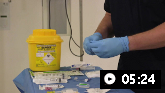
The procedure can be done quickly and easily at the bedside but must be ultrasound-guided to improve yield and safety.[7]Roberts ME, Rahman NM, Maskell NA, et al. British Thoracic Society Guideline for pleural disease. Thorax. 2023 Jul;78(suppl 3):s1-42.
https://thorax.bmj.com/content/78/Suppl_3/s1.long
Chest x-ray after thoracentesis is not routinely indicated unless air is drawn during the procedure or the patient develops symptoms, such as increased dyspnea, cough, or chest pain.[27]Petersen WG, Zimmerman R. Limited utility of chest radiograph after thoracentesis. Chest. 2000 Apr;117(4):1038-42.
http://www.ncbi.nlm.nih.gov/pubmed/10767236?tool=bestpractice.com
Establish whether the effusion is a transudate or an exudate.
Presentation with bilateral effusions or associated ascites are strongly associated with transudates.[20]Laursen CB, Clive A, Hallifax R, et al. European Respiratory Society statement on thoracic ultrasound. Eur Respir J. 2021 Mar 4;57(3):2001519.
https://erj.ersjournals.com/content/57/3/2001519.long
http://www.ncbi.nlm.nih.gov/pubmed/33033148?tool=bestpractice.com
[28]Kataoka H. Ultrasound pleural effusion sign as a useful marker for identifying heart failure worsening in established heart failure patients during follow-up. Congest Heart Fail. 2012 Sep-Oct;18(5):272-7.
https://onlinelibrary.wiley.com/doi/10.1111/j.1751-7133.2012.00285.x
http://www.ncbi.nlm.nih.gov/pubmed/22994441?tool=bestpractice.com
[29]Gurung P, Goldblatt M, Huggins JT, et al. Pleural fluid analysis and radiographic, sonographic, and echocardiographic characteristics of hepatic hydrothorax. Chest. 2011 Aug;140(2):448-53.
https://www.ncbi.nlm.nih.gov/pmc/articles/PMC3148794
http://www.ncbi.nlm.nih.gov/pubmed/21273292?tool=bestpractice.com
On TUS this makes the fluid appear anechoic, however this is not specific.[30]Sajadieh H, Afzali F, Sajadieh V, et al. Ultrasound as an alternative to aspiration for determining the nature of pleural effusion, especially in older people. Ann N Y Acad Sci. 2004 Jun;1019:585-92.
http://www.ncbi.nlm.nih.gov/pubmed/15247092?tool=bestpractice.com
In contrast, effusions which are exudates will almost always appear echogenic, complex, or both.[30]Sajadieh H, Afzali F, Sajadieh V, et al. Ultrasound as an alternative to aspiration for determining the nature of pleural effusion, especially in older people. Ann N Y Acad Sci. 2004 Jun;1019:585-92.
http://www.ncbi.nlm.nih.gov/pubmed/15247092?tool=bestpractice.com
25 to 50 mL of pleural fluid should be submitted for cytologic analysis. If only smaller volumes are available, these may be sent but clinicians should be aware of the reduced sensitivity.[7]Roberts ME, Rahman NM, Maskell NA, et al. British Thoracic Society Guideline for pleural disease. Thorax. 2023 Jul;78(suppl 3):s1-42.
https://thorax.bmj.com/content/78/Suppl_3/s1.long
The pleural lactate dehydrogenase (LDH) and protein levels, and serum LDH and protein, should be measured to determine whether the effusion is a transudate or exudate using the Light criteria (where one or more of the following suggest an exudate: pleural fluid protein divided by serum protein >0.5; pleural fluid LDH divided by serum LDH >0.6; and pleural fluid LDH >two-thirds of the upper limit of laboratory normal range for serum LDH).[12]Saguil A, Wyrick K, Hallgren J. Diagnostic approach to pleural effusion. Am Fam Physician. 2014 Jul 15;90(2):99-104.
https://www.aafp.org/afp/2014/0715/p99.html
http://www.ncbi.nlm.nih.gov/pubmed/25077579?tool=bestpractice.com
Pleural fluid cholesterol analysis has been used to distinguish exudates from transudates but is not used routinely.[31]McGrath EE, Blades Z, Anderson PB. Chylothorax: aetiology, diagnosis and therapeutic options. Respir Med. 2010 Jan;104(1):1-8.
http://www.ncbi.nlm.nih.gov/pubmed/19766473?tool=bestpractice.com
[32]McGrath EE, Warriner D, Anderson PB. The use of non-routine pleural fluid analysis in the diagnosis of pleural effusion. Respir Med. 2010 Aug;104(8):1092-100.
http://www.ncbi.nlm.nih.gov/pubmed/20392619?tool=bestpractice.com
In one meta-analysis to determine the best means for differentiating exudates from transudates, diagnosis of an exudate was most accurate with pleural fluid cholesterol >55 mg/dL or pleural fluid to serum cholesterol ratio of >0.3.[3]Wilcox ME, Chong CA, Stanbrook MB, et al. Does this patient have an exudative pleural effusion? The Rational Clinical Examination systematic review. JAMA. 2014 Jun 18;311(23):2422-31.
http://www.ncbi.nlm.nih.gov/pubmed/24938565?tool=bestpractice.com
A transudate is likely to be caused by CHF, cirrhosis, or nephrosis. An exudative effusion will require further studies. In congestive cardiac failure, diuretic therapy, which is the mainstay of treatment, can cause elevated levels of pleural fluid protein and LDH, resulting in a misclassification of pleural effusions as exudative in up to 25% of cases.[33]Romero-Candeira S, Fernández C, Martín C, et al. Influence of diuretics on the concentration of proteins and other components of pleural transudates in patients with heart failure. Am J Med. 2001 Jun 15;110(9):681-6.
http://www.ncbi.nlm.nih.gov/pubmed/11403751?tool=bestpractice.com
In cases where no cause for an exudative effusion can be identified, or congestive cardiac failure is suspected, there is some evidence that the sequential application of the pleural fluid LDH, followed by the serum-to-pleural fluid protein, and then the serum-to-protein albumin gradients may assist in diagnosis.[34]Kummerfeldt CE, Chiuzan CC, Huggins JT, et al. Improving the predictive accuracy of identifying exudative effusions. Chest. 2014 Mar 1;145(3):586-92.
http://www.ncbi.nlm.nih.gov/pubmed/24008773?tool=bestpractice.com
Pleural fluid N-terminal pro-brain natriuretic peptide (NT-pro-BNP) should be considered in patients with pleural effusions, suspected of having CHF, however this is not superior to serum NT-pro-BNP so should not be used routinely.[7]Roberts ME, Rahman NM, Maskell NA, et al. British Thoracic Society Guideline for pleural disease. Thorax. 2023 Jul;78(suppl 3):s1-42.
https://thorax.bmj.com/content/78/Suppl_3/s1.long
[35]Zhou Q, Ye ZJ, Su Y, et al. Diagnostic value of N-terminal pro-brain natriuretic peptide for pleural effusion due to heart failure: a meta-analysis. Heart. 2010 Aug;96(15):1207-11.
http://www.ncbi.nlm.nih.gov/pubmed/20511623?tool=bestpractice.com
A laboratory cutoff value of 1500 picograms/mL is commonly used.[36]Janda S, Swiston J. Diagnostic accuracy of pleural fluid NT-pro-BNP for pleural effusions of cardiac origin: a systematic review and meta-analysis. BMC Pulm Med. 2010 Nov 20;10:58.
https://bmcpulmmed.biomedcentral.com/articles/10.1186/1471-2466-10-58
http://www.ncbi.nlm.nih.gov/pubmed/21092122?tool=bestpractice.com
Chylothorax
Chyle (a sterile, odorless, alkaline, milky fluid) is sometimes difficult to differentiate from empyema, but if chylothorax is suspected, lipid analysis of the pleural fluid should be performed. The presence of chylomicrons on microscopy confirms a chylothorax and a high triglyceride level, usually >110 mg/dL is diagnostic.[37]Majdalany BS, Murrey DA Jr, et al; Expert Panel on Vascular Imaging and Interventional Radiology. ACR appropriateness criteria®: chylothorax treatment planning. J Am Coll Radiol. 2017 May;14(5s):S118-26.
https://www.jacr.org/article/S1546-1440(17)30205-3/fulltext
http://www.ncbi.nlm.nih.gov/pubmed/28473067?tool=bestpractice.com
Chylothorax can usually be excluded if the triglyceride level is <50 mg/dL.[16]McGrath EE, Anderson PB. Diagnosis of pleural effusion: a systematic approach. Am J Crit Care. 2011 Mar;20(2):119-27.
http://www.ncbi.nlm.nih.gov/pubmed/21362716?tool=bestpractice.com
Blood tests and cultures
Total and differential cell count, glucose, pH, and cytology are recommended; routine cultures are not useful or cost effective, but if a parapneumonic effusion is suspected or there is frank pus, then this is a valuable test.[38]Jimenez D, Diaz G, Garcia-Rull S, et al. Routine use of pleural fluid cultures. Are they indicated? Limited yield, minimal impact on treatment decisions. Respir Med. 2006 Nov;100(11):2048-52.
http://www.ncbi.nlm.nih.gov/pubmed/16584878?tool=bestpractice.com
Clinicians should be aware that using pleural cytology for the diagnosis of malignant pleural effusion can be unreliable, as it has a low sensitivity of 58%.[39]Kassirian S, Hinton SN, Cuninghame S, et al. Diagnostic sensitivity of pleural fluid cytology in malignant pleural effusions: systematic review and meta-analysis. Thorax. 2023 Jan;78(1):32-40.
http://www.ncbi.nlm.nih.gov/pubmed/35110369?tool=bestpractice.com
Pleural fluid should be inoculated into aerobic and anaerobic blood culture bottles at the same time as standard culture as this increases microbial yield.[40]Menzies SM, Rahman NM, Wrightson JM, et al. Blood culture bottle culture of pleural fluid in pleural infection. Thorax. 2011 Aug;66(8):658-62.
http://www.ncbi.nlm.nih.gov/pubmed/21459855?tool=bestpractice.com
Pleural fluid pH <7.20 is highly suggestive of pleural infection or complex parapneumonic effusion (CPPE); clear pleural fluid for pH can be collected anaerobically with heparin and measured in a blood gas analyzer.[41]Bhatnagar R, Maskell N. The modern diagnosis and management of pleural effusions. BMJ. 2015 Sep 8;351:h4520.
http://www.ncbi.nlm.nih.gov/pubmed/26350935?tool=bestpractice.com
(Avoid putting turbid fluid or frank pus through the analyzer. Note that manufacturers of blood gas analyzers may void their warranty if any fluid other than blood is processed through the machine.) In the absence of readily available pleural fluid pH measurement, an initial pleural fluid glucose <3.3 mmol/L may be used as an indicator of high probability of CPPE/pleural infection and can be used to inform decision to insert an intercostal drain in the appropriate clinical context.[7]Roberts ME, Rahman NM, Maskell NA, et al. British Thoracic Society Guideline for pleural disease. Thorax. 2023 Jul;78(suppl 3):s1-42.
https://thorax.bmj.com/content/78/Suppl_3/s1.long
If the cell count is predominantly lymphocyte-dominant, then a test for markers of tuberculosis (TB) in the pleural fluid, such as adenosine deaminase, should be requested.[42]Liang QL, Shi HZ, Wang K, et al. Diagnostic accuracy of adenosine deaminase in tuberculous pleurisy: a meta-analysis. Respir Med. 2008 May;102(5):744-54.
http://www.ncbi.nlm.nih.gov/pubmed/18222681?tool=bestpractice.com
[43]Aggarwal AN, Agarwal R, Sehgal IS, et al. Meta-analysis of Indian studies evaluating adenosine deaminase for diagnosing tuberculous pleural effusion. Int J Tuberc Lung Dis. 2016 Oct;20(10):1386-91.
https://www.ingentaconnect.com/content/iuatld/ijtld/2016/00000020/00000010/art00023%3bj
http://www.ncbi.nlm.nih.gov/pubmed/27725052?tool=bestpractice.com
Eosinophilic pleural effusions (defined as >10% of pleural white cells) account for 10% of exudates and are nonspecific. Causes include malignancy (26%), idiopathic (25%), related to pleural air or blood (13%), parapneumonic (13%), and TB (7%), along with other less common miscellaneous causes. Likelihood of malignancy is inversely correlated with eosinophil count.[44]Oba Y, Abu-Salah T. The prevalence and diagnostic significance of eosinophilic pleural effusions: a meta-analysis and systematic review. Respiration. 2012;83(3):198-208.
http://www.ncbi.nlm.nih.gov/pubmed/21576924?tool=bestpractice.com
Interferon-gamma measurement in pleural fluid is sensitive and specific for the diagnosis of tuberculous pleurisy.[45]Jiang J, Shi HZ, Liang QL, et al. Diagnostic value of interferon-gamma in tuberculous pleurisy: a metaanalysis. Chest. 2007;131:1133-1141.
http://www.ncbi.nlm.nih.gov/pubmed/17426220?tool=bestpractice.com
[46]Kalantri Y, Hemvani N, Chitnis DS. Evaluation of real-time polymerase chain reaction, interferon-gamma, adenosine deaminase, and immunoglobulin A for the efficient diagnosis of pleural tuberculosis. Int J Infect Dis. 2011 Apr;15(4):e226-31.
http://www.ncbi.nlm.nih.gov/pubmed/21227729?tool=bestpractice.com
[47]Mollo B, Jouveshomme S, Philippart F, et al. Biological markers in the diagnosis of tuberculous pleural effusion [in French]. Ann Biol Clin (Paris). 2017 Feb 1;75(1):19-27.
http://www.ncbi.nlm.nih.gov/pubmed/28057604?tool=bestpractice.com
T-cell gamma interferon assays (IGRA) on blood or pleural fluid are not sensitive or specific enough to be clinically useful for diagnosis of TB pleurisy.[48]Zhou Q, Chen YQ, Qin SM, et al. Diagnostic accuracy of T-cell interferon-γ release assays in tuberculous pleurisy: a meta-analysis. Respirology. 2011;16:473-480.
http://www.ncbi.nlm.nih.gov/pubmed/21299686?tool=bestpractice.com
Subsequent imaging studies
Thoracic computed tomography (CT) scans are useful to define the size and location of the effusion, show loculations, and identify additional pathology that requires further investigation (e.g., a lung mass or pleural thickening). A simple CT-scan scoring system to distinguish malignant effusions from benign effusions has been validated in two small populations.[49]Porcel JM, Pardina M, Bielsa S, et al. Derivation and validation of a CT scan scoring system for discriminating malignant from benign pleural effusions. Chest. 2015 Feb;147(2):513-9.
http://www.ncbi.nlm.nih.gov/pubmed/25255186?tool=bestpractice.com
If there is suspicion of TB, thoracic CT is indicated for detecting subtle parenchymal changes, as well as mediastinal lymphadenopathy, which may represent a target for transbronchial nodal aspiration via bronchoscopy. CT follow-up should also be considered for patients presenting with pleural infection to exclude occult malignancy if there are ongoing symptoms, or other clinically concerning features.[7]Roberts ME, Rahman NM, Maskell NA, et al. British Thoracic Society Guideline for pleural disease. Thorax. 2023 Jul;78(suppl 3):s1-42.
https://thorax.bmj.com/content/78/Suppl_3/s1.long
Magnetic resonance imaging (MRI), which provides better imaging of soft tissues than CT, can reveal tumor invasion of the chest wall or diaphragm, and can distinguish between benign and malignant effusions (using differences in signal intensity). MRI is, however, not routinely indicated in investigation of pleural effusion, and first-line cross-sectional imaging should be CT.[17]Expert Panel on Thoracic Imaging, Morris MF, Henry TS, et al. ACR Appropriateness Criteria® Workup of pleural effusion or pleural Disease. J Am Coll Radiol. 2024 Jun;21(6):S343-52.
http://www.ncbi.nlm.nih.gov/pubmed/38823955?tool=bestpractice.com
[50]Heffner JE, Klein JS. Recent advances in the diagnosis and management of malignant pleural effusions. Mayo Clin Proc. 2008 Feb;83(2):235-50.
http://www.ncbi.nlm.nih.gov/pubmed/18241636?tool=bestpractice.com
If no cause is established, pulmonary embolus should be ruled out by further pulmonary imaging, such as a helical CT scan. Pleural effusions due to pulmonary emboli are usually small and unilateral exudates.[51]Porcel JM, Light RW. Pleural effusions due to pulmonary embolism. Curr Opin Pulm Med. 2008 Jul;14(4):337-42.
http://www.ncbi.nlm.nih.gov/pubmed/18520269?tool=bestpractice.com
Bronchoscopy, thoracoscopy, and tissue biopsy
Bronchoscopy is not routinely indicated in the investigation of pleural effusion. However, if the cause of an exudative effusion cannot be established, bronchoscopy can be used to exclude small malignant endobronchial lesions. Bronchoscopy should be performed after pleural fluid drainage to ensure optimum diagnostic conditions.
Thoracoscopy for diagnostic purposes is indicated if the patient is not improving, the cause of the effusion is unknown, TB is suspected, or cytology is negative when pleural malignancy is suspected. If malignancy is confirmed, then thoracoscopy can also be therapeutic and complication rates tend to be low.[12]Saguil A, Wyrick K, Hallgren J. Diagnostic approach to pleural effusion. Am Fam Physician. 2014 Jul 15;90(2):99-104.
https://www.aafp.org/afp/2014/0715/p99.html
http://www.ncbi.nlm.nih.gov/pubmed/25077579?tool=bestpractice.com
[52]Martinez-Zayas G, Molina S, Ost DE. Sensitivity and complications of thoracentesis and thoracoscopy: a meta-analysis. Eur Respir Rev. 2022 Dec 21;31(166):220053.
https://err.ersjournals.com/content/31/166/220053.long
http://www.ncbi.nlm.nih.gov/pubmed/36543349?tool=bestpractice.com
Thoracoscopy is traditionally carried out by surgeons, but medical rigid or semi-rigid thoracoscopy is a safe, simple, and accurate alternative.[53]Mohan AC, Chandra S, Agarwal D, et al. Utility of semirigid thoracoscopy in the diagnosis of pleural effusions: a systematic review. J Bronchology Interv Pulmonol. 2010 Jul;17(3):195-201.
http://www.ncbi.nlm.nih.gov/pubmed/23168883?tool=bestpractice.com
One meta-analysis of the usefulness of semi-rigid thoracoscopy in undiagnosed exudative pleural effusions (following thoracentesis with or without blind pleural biopsy) found a pooled sensitivity of 91%, with a specificity of 100%.[54]Agarwal R, Aggarwal AN, Gupta D. Diagnostic accuracy and safety of semirigid thoracoscopy in exudative pleural effusions: a meta-analysis. Chest. 2013 Dec;144(6):1857-67.
http://www.ncbi.nlm.nih.gov/pubmed/23928984?tool=bestpractice.com
Closed pleural biopsy is used after ultrasound exam in undiagnosed exudative effusions for suspected TB or malignancy when thoracoscopy is not available. The European Respiratory Society (ERS) and the European Association for Cardio-Thoracic Surgery (EACTS) suggest that biopsy is the definitive test for diagnosis and treatment planning of malignant pleural effusion.[55]Bibby AC, Dorn P, Psallidas I, et al. ERS/EACTS statement on the management of malignant pleural effusions. Eur Respir J. 2018 Jul;52(1):.
https://www.doi.org/10.1183/13993003.00349-2018
http://www.ncbi.nlm.nih.gov/pubmed/30054348?tool=bestpractice.com
Nonimage guided pleural biopsies should not be conducted.[7]Roberts ME, Rahman NM, Maskell NA, et al. British Thoracic Society Guideline for pleural disease. Thorax. 2023 Jul;78(suppl 3):s1-42.
https://thorax.bmj.com/content/78/Suppl_3/s1.long
Procalcitonin
Procalcitonin is now commonly used as a biomarker for the diagnosis of bacterial infections.[56]Schuetz P, Wirz Y, Sager R, et al. Procalcitonin to initiate or discontinue antibiotics in acute respiratory tract infections. Cochrane Database Syst Rev. 2017 Oct 12;10:CD007498.
https://www.doi.org/10.1002/14651858.CD007498.pub3
http://www.ncbi.nlm.nih.gov/pubmed/29025194?tool=bestpractice.com
[57]Huang DT, Yealy DM, Filbin MR, et al. Procalcitonin-guided use of antibiotics for lower respiratory tract infection. N Engl J Med. 2018 Jul 19;379(3):236-249.
https://www.doi.org/10.1056/NEJMoa1802670
http://www.ncbi.nlm.nih.gov/pubmed/29781385?tool=bestpractice.com
[58]Yancey JR, Nelson MD, Whalen NJ. Procalcitonin for diagnosis, risk assessment, and prognosis of respiratory tract infections. Am Fam Physician. 2022 Sep;106(3):333-4. Higher levels of procalcitonin have been detected in severe bacterial infections.[56]Schuetz P, Wirz Y, Sager R, et al. Procalcitonin to initiate or discontinue antibiotics in acute respiratory tract infections. Cochrane Database Syst Rev. 2017 Oct 12;10:CD007498.
https://www.doi.org/10.1002/14651858.CD007498.pub3
http://www.ncbi.nlm.nih.gov/pubmed/29025194?tool=bestpractice.com
[58]Yancey JR, Nelson MD, Whalen NJ. Procalcitonin for diagnosis, risk assessment, and prognosis of respiratory tract infections. Am Fam Physician. 2022 Sep;106(3):333-4. Do not order procalcitonin without an established, evidence-based protocol.[59]American Society for Clinical Pathology. Thirty five things physicians and patients should question. Choosing Wisely, an initiative of the ABIM Foundation. 2021 [internet publication].
https://web.archive.org/web/20230316185857/https://www.choosingwisely.org/societies/american-society-for-clinical-pathology
It may have a function in guiding when to use antibiotics for the treatment of lower respiratory tract infection; however, this is unclear. A Cochrane review of the use of procalcitonin to guide initiation and duration of antibiotic treatment in people with acute respiratory tract infections found it lowered the risk of mortality, and led to lower antibiotic consumption and lower risk for antibiotic-related side effects in all patients.[56]Schuetz P, Wirz Y, Sager R, et al. Procalcitonin to initiate or discontinue antibiotics in acute respiratory tract infections. Cochrane Database Syst Rev. 2017 Oct 12;10:CD007498.
https://www.doi.org/10.1002/14651858.CD007498.pub3
http://www.ncbi.nlm.nih.gov/pubmed/29025194?tool=bestpractice.com
Further research is required to establish its use in clinical practice. In a separate analysis of 1656 patients, 826 were randomly assigned to a group where the decision on whether to provide antibiotics was based on the results of a procalcitonin assay (830 patients were given usual care).[57]Huang DT, Yealy DM, Filbin MR, et al. Procalcitonin-guided use of antibiotics for lower respiratory tract infection. N Engl J Med. 2018 Jul 19;379(3):236-249.
https://www.doi.org/10.1056/NEJMoa1802670
http://www.ncbi.nlm.nih.gov/pubmed/29781385?tool=bestpractice.com
The assay results did not result in less use of antibiotics. There was no significant difference between the procalcitonin group and the usual-care group in antibiotic-days (mean 4.2 and 4.3 days, respectively; difference −0.05 days; 95% CI −0.6 to +0.5; P = 0.87) or the proportion of patients with adverse outcomes (11.7% [96 patients] and 13.1% [109 patients]; difference −1.5 percentage points; 95% CI, −4.6 to +1.7; P <0.001 for non-inferiority) within 30 days.