Approach
Although the causes are numerous, a thorough history and physical exam helps elucidate the diagnosis in most patients.
History
The primary purpose of the initial encounter is to evaluate whether the symptoms suggest a more serious underlying condition.[5] Musculoskeletal lower back pain can be diagnosed clinically from the history and physical examination. It is an exclusion diagnosis, therefore the clinician must eliminate specific lower back pain causes of neurologic compromise, neoplasia, inflammatory arthritis, fracture, or referred pain from other locations or organ systems.
Red flags
Red flag signs and symptoms warranting additional diagnostic imaging and emergent referral to a spine specialist for ongoing management may include:[29][30][31][32][33][34][35]
Saddle anesthesia
Sphincter disturbance (bladder or bowel dysfunction e.g. acute urinary retention, new onset urinary or fecal incontinence, loss of anal sphincter tone)
Profound or progressive neurologic deficit
History of malignancy with new onset back pain
Systemic ailments, including fever, chills, night sweats, and/or unexplained weight loss (infection/malignancy)
Intravenous drug use
Urinary tract infection
Immunosuppression, including prolonged corticosteroid use or other immunosuppressive therapies
Trauma (including minor trauma in older adults)
Presence of contusion or abrasions over the spine
History of osteoporosis
Pain that is refractory to conservative management
Thoracic pain
Non-mechanical pain (i.e., systemic or referred causes of pain). Pain at rest and at night suggests a non-mechanical cause
Age >50 years.
Red flag signs and symptoms vary between guidelines.[36] Most guidelines endorse the red flags of history of malignancy, unexpected weight loss, significant trauma, prolonged corticosteroid use, fever and HIV.[5]
Patients should be questioned regarding prior back pain episodes and treatments, onset, duration, location, radiation, character, aggravating and relieving factors, and severity. Musculoskeletal lower back pain may be dull, gnawing, tearing, burning, electric and/or associated with muscle spasms. Associated sciatica (radicular pain in the distribution of the sciatic nerve) usually presents as unilateral pain radiating from the buttock to below the knee.[51] Sciatic pain often has aching and sharp qualities and the patient may report associated weakness, numbness or paresthesia.[52] Pain from spinal stenosis is typically worse with standing and lumbar extension and is relieved by sitting or forward flexion. Postural changes trigger neurogenic claudication, with symptoms of pain, weakness, numbness or tingling in the lower back, legs and buttocks.[19]
Further inquiry into functional, occupational, social, and psychiatric history should be sought to address risk factors, including obesity, occupational risks, smoking, and psychosocial stressors.[53]
Maladaptive pain coping behaviors, nonorganic signs, functional impairment, poor general health status, and psychiatric comorbidities are risk factors for developing chronic disabling low back pain.[9]
Symptoms of severe compression of the cauda equina or spinal cord
Bowel or bladder dysfunction, bilateral sciatica, and saddle anesthesia may be symptoms of severe compression of the cauda equina. The etiology is usually a large central herniated disk or a pathologic or traumatic fracture. Acute spinal cord compression may present with sensory symptoms of altered sensation below a certain level or hemisensory loss; motor symptoms of hemiplegia/hemiparesis, paraplegia/paraparesis or tetraplegia/tetraparesis; and/or autonomic symptoms including constipation and urinary retention. The etiology may be spine trauma, vertebral compression fracture, intervertebral disc herniation, primary or metastatic spinal tumor, or infection.
A complete history and physical exam should identify impending neurologic compromise and the need for emergent referral to a spinal surgeon.
Infections
Important infections to consider are spinal epidural abscess, osteomyelitis of the spine and discitis. Spinal epidural abscess can present with fever, back or neck pain, and neurologic deficits. However, this triad of symptoms is only present in 10% to 15% of cases and therefore having a low threshold for considering this diagnosis in patients at risk is crucial.[27]
Risk factors for epidural abscess include diabetes mellitus, intravenous drug use, an immunocompromised state, recent spinal surgery or trauma, presence of indwelling spinal catheter, pre-existing infection (in contiguous tissue or distant infection causing bacteremia), dialysis and alcohol misuse.[27]
Osteomyelitis and discitis of the lumbar vertebrae can present with low back pain and a low grade fever.[54] It may be associated with intravenous drug use, lower extremity and hip infections, and tuberculosis, due to the anatomy of the Batson venous plexus.[55][56]
It is important to consider vertebral osteomyelitis and discitis in older adults with back pain and urinary tract symptoms as the urinary tract may be a source of infection from gram-negative organisms.[54]
Herpes zoster (shingles) affecting a truncal dermatome may present with back pain. There is typically a prodrome of burning, stinging or stabbing pain, followed by a vesicular rash which does not cross the midline.
Spinal metastasis
Metastasis to the spine needs to be excluded, especially in patients with a current diagnosis of cancer, a history of cancer, or suspected cancer.[39] Breast, prostate, and lung cancer are responsible for more than 80% of cases of metastatic bone disease and the spine is the most common site of bone metastasis.[57]
Spinal compression fractures
Should be considered in patients who are at risk of osteoporotic disease. Older people and those on long-term corticosteroid therapy are particularly at risk.
Many osteoporotic spinal compression fractures are asymptomatic with no recognized trauma and are identified incidentally on radiographs while investigating other pathologies. However, some patients can present with acute onset back pain from minor trauma such as coughing or sneezing. The pain often disturbs sleep, is aggravated by movement and can radiate bilaterally to the abdomen.
Inflammatory spondyloarthropathy
Back pain due to inflammatory spondyloarthropathy more commonly starts before the age of 35 years. There may be a family history of psoriasis or spondyloarthritis, or a history of recent genitourinary infection. Other features suggesting spondyloarthritis include waking due to pain in the second half of the night, buttock pain, improvement with movement and improvement within 48 hours of taking nonsteroidal anti-inflammatory drugs.[28]
Musculoskeletal symptoms apart from chronic back pain include dactylitis and enthesitis. Extra-articular symptoms include uveitis and psoriasis (including psoriatic nail symptoms).
It is important to be aware that the diagnosis of spondyloarthritis is sometimes missed or delayed.[28] Prompt referral to a rheumatologist is therefore essential for further diagnostic tests and appropriate management.
Nonspecific causes of back pain
It is important to identify any less serious or nonspecific causes of back pain to ensure appropriate management and maximum quality of life for these patients. While a specific cause of low back pain can rarely be identified, the most prevalent type is mechanical nonspecific low back pain (exacerbated by movement and relieved by rest).[6][58]
Risk stratification tools can be used (e.g., the STarT Back risk assessment tool) at first point of contact for each new episode of low back pain, with or without sciatica, to inform shared decision-making about stratified management.[7]
Physical exam
Perform a focused musculoskeletal and neurologic exam.
A musculoskeletal exam consists of the following:
Inspection
Looking for obvious deformity (e.g., in fractures) and abnormal curvature (scoliosis, kyphosis, lordosis), which can create pain in a minority of cases. This should prompt the clinician to order radiographs to document baseline curvature and orthopedic referral for moderate to severe cases. Patients with spondylolisthesis may have an exaggerated lordosis and heart-shaped buttock.
Palpation
Palpation of the spinous processes and musculature: to localize any tenderness and to detect the presence of a midline "step-off" of the spinous processes that may indicate spondylolisthesis.[59] Tenderness on palpation over the sacroiliac joint may indicate sacroiliitis. Localized tenderness, present particularly with percussion, can indicate vertebral discitis or osteomyelitis.
Movement
Active and passive range of motion (ROM) assessment:[60] patients are asked in standing position to actively flex, extend, and laterally bend as far as they can. Pain on flexion that radiates to the leg suggests disk herniation with impingement on a nerve root; pain on extension can suggest either facet arthropathy or spinal stenosis. Greatly restricted ROM in a younger patient may suggest ankylosing spondylitis. Normal values for ROM vary between studies and decline in older age.[61] One study of asymptomatic subjects investigated the ROM of the lumbar spine during 15 activities of daily living. The median ROM used was 9 degrees for flexion/extension, 6 degrees for lateral bending and 5 degrees for rotation.[62]
Every patient with lower back pain should have a hip examination. The distribution of pain from hip disorders is similar to the distribution of pain from lumbar spine disorders.[63] Hip and spine disorders may also coexist. Passive ROM of each hip should be assessed with the patient lying supine. There should be normally 130° of flexion, extension to 15° beyond neutral, and approximately 45° of internal and external rotation.[64] Pain in any of these motions suggests hip pathology and hip x-rays should be obtained.
Patient gait and ability to walk should also be observed.
Provocative tests for lumbar disk herniation
A number of provocative tests may be performed.
The straight-leg raise or contralateral straight-leg raise test which, when positive, indicates a possible herniated nucleus pulposus (HNP). A straight-leg raise is performed with the patient supine and the hip flexed gradually with the knee extended. Pain that is reproduced below 60° of hip flexion on the ipsilateral side is considered a positive straight-leg raise and is more sensitive. Reproduced pain on the contralateral side indicates a positive contralateral straight-leg raise and is more specific. Pain that occurs above 60° is usually secondary to hamstring tightness.[65][66][67] The pain associated with HNP is usually worse in the leg than in the back, with pain radiating to the lower extremity in a dermatomal distribution. However, the absence of a positive straight-leg raise test and dermatomal pain does not exclude disk herniation.[68]
The femoral stretch or contralateral femoral stretch tests can be used to assess for upper lumbar disk herniation. The test is typically performed with the patient prone. The knee is flexed, and then the leg is extended. If it reproduces the leg pain, it is considered positive.[69]
Provocative tests for sacroiliac joint (SIJ) pain
Several tests exist to evaluate the SIJ. Three or more positive tests indicate that the SIJ is likely to be the pain generator.[70][71][72]
The thigh thrust test is the most sensitive test (sensitivity 88%).[71] The patient lies supine with the contralateral leg extended. The examiner flexes the ipsilateral hip to 50 degrees and the knee remains relaxed. The examiner encloses the knee and slightly adducts the femur, then applies a graded force through the long axis of the femur.[72]
The distraction test is the most specific (specificity 81%).[71] With the patient lying supine, the examiner applies dorsal and lateral pressure to the anterior superior iliac spines.[72]
The Gaenslen test is performed by asking the patient to lie supine with the symptomatic leg hanging over the edge of the exam table. The nonsymptomatic leg is flexed at the hip and knee. A rotational force is then applied across the sacroiliac joint by applying pressure to the flexed knee outwards, while simultaneously applying pressure downwards on the affected sacroiliac joint with the other hand.
The pelvic compression test is performed by applying compression across the sacroiliac joints while the patient lies on their side.
The FABER test is performed by asking the patient to bend their knee on the affected side and externally rotate at the hip, resting the foot across the opposite knee, so creating the shape of the number 4 with the legs. Pressure is then applied downwards on the knee on the affected side, while simultaneously using the other hand to apply pressure downwards on the opposite iliac crest.
Provocative test for spondylosis
The one-legged hyperextension test can be performed if the diagnosis of active spondylosis is suspected, but is not sufficiently sensitive to exclude the diagnosis.[73] The patient stands on the affected leg and lifts the unaffected leg, slightly flexing the hip and flexing the knee to 80 degrees. The patient then actively extends their lumbar spine. The test is positive if this action reproduces their pain.[73]
Neurologic exam
A thorough neurologic exam should follow. Specific neurologic deficits, such as weakness, spasticity, or hyper/hyporeflexia, should be noted. These suggest more profound neurologic compression; prompt referral to a spinal surgeon is indicated for further evaluation and management.[33] Decreased rectal tone is an important exam finding, as it suggests sacral root encroachment from significant intraspinal compression such as cauda equina.[33]
[Figure caption and citation for the preceding image starts]: Sensory dermatomes of the lower back and legCreated by BMJ Knowledge Centre [Citation ends].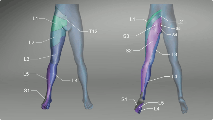
Vascular exam
A vascular exam is important in differentiating vascular versus neurogenic claudication. Vascular claudication typically worsens with ambulation in any position and is relieved immediately by rest. Neurogenic claudication worsens with ambulation in an extended posture and improves with forward flexion of the lumbar spine.[19] Patients with claudication may have concomitant vascular and spinal pathologies. Diminished pulses and mottled, thin, shiny skin are signs of peripheral arterial disease.
Other features that may aid diagnosis
A general physical exam may detect other features to aid diagnosis. For instance, when pain is referred:
Patients with abdominal aortic aneurysm may have a detectable pulsatile abdominal mass
Patients with peptic ulcer may have epigastric tenderness and melena on rectal exam
Patients with renal colic or pyelonephritis may have flank or costovertebral tenderness
Patients with pancreatitis may have a fever and tenderness/guarding of abdomen.
Patients with herpes zoster (shingles) usually have a characteristic vesicular rash in the affected dermatome.
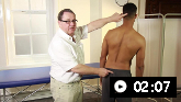
How to perform an inspection examination of the back, including inspection of gait and posture
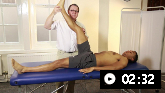
A primary care physician demonstrates how to perform a physical examination of the back
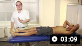
A primary care physician shows how to perform a neurological examination of the back
Laboratory tests
Routine laboratory studies are not necessary in the evaluation of back pain unless the physician is concerned about the possibility of malignancy, infection or inflammatory arthritis. In these cases complete blood count (CBC), erythrocyte sedimentation rate (ESR), and C-reactive protein (CRP) are typically obtained. Blood cultures should be taken if infection is suspected.
CBC, ESR and CRP are normal in cases of mechanical back pain. Though nonspecific abnormal values may indicate malignancy, infection or inflammatory arthritis and should prompt further investigation of the cause of back pain.
A urinalysis and urine culture should be ordered when considering the possibility of pyelonephritis or renal colic.
Imaging
Most patients with low back pain, with or without sciatica, do not routinely require imaging when presenting in a nonspecialist setting.[7][Evidence C]
If there are no red flags or high-risk features, they should be reassured that their symptoms will respond to conservative treatment.
If symptoms are progressive or persist longer than 6 weeks, imaging should be obtained if the result will change clinical management, for example if the patient is a candidate for surgery or intervention or if there is diagnostic uncertainty.[7][29]
Degenerative and disk abnormalities are found in many asymptomatic patients, causing overdiagnosis and unwarranted patient anxiety.[29][66][74][75][76][77] Degenerative findings do not necessarily equate to symptomatic lesions.[78][79] Furthermore, there is no correlation between severity of symptoms and findings on MRI.[80] Therefore, imaging studies should be ordered after discussion with a spinal surgeon or by the spinal surgeon in most cases to prevent ordering of unnecessary tests.
When imaging is indicated, magnetic resonance imaging is usually the diagnostic modality of choice.[29]
Plain radiographs
May be indicated when there is a suspicion of a vertebral fracture: for example, in patients with low velocity trauma, especially minor trauma in older adults, and those with osteoporosis or chronic corticosteroid use.[29][81]
Plain radiographs are performed in people with suspected sacroiliitis to rule out other causes of pain, but there are no pathognomonic findings specific to sacroiliac joint pain.[82]
If plain radiographs are obtained following thoracolumbar spinal trauma, anteroposterior and lateral views are required. A "swimmer’s lateral" view should be obtained if the shoulders obscure the upper thoracic spine.[31] However, computed tomography (CT) is the diagnostic modality of choice in these patients.[31]
Magnetic resonance imaging (MRI)
Urgent MRI is indicated if neurologic compromise is present, or if infection or tumor is suspected.[29][38][39]
If a patient has metal in their body or is unable to undergo an MRI, a CT myelogram is usually warranted.[29]
MRI scan of the sacroiliac joints is not generally required, but is recommended when inflammatory spondylitis is suspected as the cause of back pain but the clinical exam and plain radiograph has not established the diagnosis.[82]
MRI is sometimes used to diagnose spondylosis or spondylolisthesis.[83]
Computed tomography (CT)
CT imaging of the thoracolumbar spine is the preferred test following spinal trauma for patients with midline tenderness, a high energy mechanism of injury, or those who are >60 years with a mechanism of injury consistent with thoracolumbar spine injury. CT may also be required in patients who cannot be examined due to intoxication, Glasgow Coma Score <15‚ or a distracting injury.[31] CT may also be indicated if spinal metastases or metastatic spinal cord compression is suspected, and MRI is contraindicated.[39]
Neurologic compromise, gross spinal deformities or manual step off on spinal palpation also warrant CT. CT has a higher sensitivity for detecting fractures of the thoracolumbar spine than plain radiographs and also identifies soft tissue injuries that often accompany spinal fractures.[31][40]
Up to 20% of patients with spinal injuries have a second, noncontiguous injury, therefore imaging of the entire spine is recommended.[31]
Bone scintigraphy
Bone scintigraphy may be performed to detect vertebral compression fractures in patients with contraindications to magnetic resonance imaging.
Bone scan with single-photon emission CT (SPECT) or SPECT/CT is usually not used for initial imaging but can be useful for radiographically occult fractures and to evaluate acuity of vertebral fracture.[29] It is also used to detect bone metastases.[29]
Bone scintigraphy with single photon emission computed tomography is the gold standard for diagnosis of spondylolisthesis.[73]
Further investigations
Diagnosis of sacroiliitis as the cause of back pain is usually made by physical exam. However, further investigations include diagnostic local anesthetic block injection of the sacroiliac joint under C-arm fluoroscopic guidance that, when positive, provides corresponding pain relief.[72]
Other investigations, including laboratory, imaging, and endoscopic tests, are used when pain is suspected to be nonspine related (e.g., referred from intra/retroperitoneal pathologies). See Differential diagnosis.
Use of this content is subject to our disclaimer