Images and videos
Images
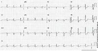


Non-ST-elevation myocardial infarction
ECG showing ST depression
From the personal collection of Dr Syed W. Yusuf and Dr Iyad N. Daher, Department of Cardiology, University of Texas, Houston; used with permission
See this image in context in the following section/s:
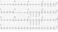
Non-ST-elevation myocardial infarction
ECG showing ST depression
From the personal collection of Dr Syed W. Yusuf and Dr Iyad N. Daher, Department of Cardiology, University of Texas, Houston; used with permission
See this image in context in the following section/s:
Non-ST-elevation myocardial infarction
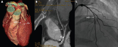
64-slice CT angiography of a patient with stable angina showing 3D reconstruction (A), curved reformatted images (B) and confirmation of a high-grade lesion on conventional angiography (C). The arrowheads show calcified plaques. Dx= diagnosis
From: Schussler JM and Grayburn PA. Heart. 2007 Mar;93(3):290-7
See this image in context in the following section/s:
Non-ST-elevation myocardial infarction
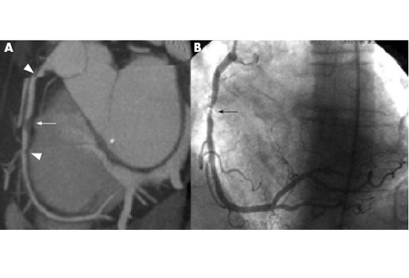
64-slice CT angiography (A) and conventional angiography (B) showing a high-grade lesion in the mid-right coronary artery, indicated by the arrows. The arrowheads show artifacts that may be mistaken for lesions
From: Schussler JM and Grayburn PA. Heart. 2007 Mar;93(3):290-7
See this image in context in the following section/s:
Use of this content is subject to our disclaimer