Patients presenting with a ruptured aneurysm require emergent repair.
For patients with symptomatic aortic aneurysms, repair is indicated regardless of diameter.[5]Isselbacher EM, Preventza O, Hamilton Black J 3rd, et al. 2022 ACC/AHA guideline for the diagnosis and management of aortic disease: a report of the American Heart Association/American College of Cardiology Joint Committee on Clinical Practice Guidelines. Circulation. 2022 Dec 13;146(24):e334-e482.
https://www.doi.org/10.1161/CIR.0000000000001106
http://www.ncbi.nlm.nih.gov/pubmed/36322642?tool=bestpractice.com
[78]National Institute for Health and Care Excellence. Abdominal aortic aneurysm: diagnosis and management. Mar 2020 [internet publication].
https://www.nice.org.uk/guidance/ng156
For asymptomatic AAA detected as an incidental finding, surveillance is preferred to repair until the theoretical risk of rupture exceeds the estimated risk of operative mortality. Generally, repair is indicated in patients with large asymptomatic AAA (e.g., with a diameter exceeding 5.5 cm in men or 5.0 cm in women in the US, although treatment decisions based on greater size may differ in other countries).[1]Dehlin JM, Upchurch GR. Management of abdominal aortic aneurysms. Curr Treat Options Cardiovasc Med. 2005 Jun;7(2):119-30.
http://www.ncbi.nlm.nih.gov/pubmed/15935120?tool=bestpractice.com
[5]Isselbacher EM, Preventza O, Hamilton Black J 3rd, et al. 2022 ACC/AHA guideline for the diagnosis and management of aortic disease: a report of the American Heart Association/American College of Cardiology Joint Committee on Clinical Practice Guidelines. Circulation. 2022 Dec 13;146(24):e334-e482.
https://www.doi.org/10.1161/CIR.0000000000001106
http://www.ncbi.nlm.nih.gov/pubmed/36322642?tool=bestpractice.com
[76]Chaikof EL, Dalman RL, Eskandari MK, et al. The Society for Vascular Surgery practice guidelines on the care of patients with an abdominal aortic aneurysm. J Vasc Surg. 2018 Jan;67(1):2-77.e2.
https://www.jvascsurg.org/article/S0741-5214(17)32369-8/fulltext
http://www.ncbi.nlm.nih.gov/pubmed/29268916?tool=bestpractice.com
[78]National Institute for Health and Care Excellence. Abdominal aortic aneurysm: diagnosis and management. Mar 2020 [internet publication].
https://www.nice.org.uk/guidance/ng156
[103]Lederle FA, Wilson SE, Johnson GR, et al; Aneurysm Detection and Management Veterans Affairs Cooperative Study Group. Immediate repair compared with surveillance of small abdominal aortic aneurysms. N Engl J Med. 2002 May 9;346(19):1437-44.
http://www.ncbi.nlm.nih.gov/pubmed/12000813?tool=bestpractice.com
[104]UK Small Aneurysm Trial Participants. Mortality results for randomized controlled trial of early elective surgery or ultrasonographic surveillance for small abdominal aortic aneurysms. Lancet. 1998 Nov 21;352(9141):1649-55.
http://www.ncbi.nlm.nih.gov/pubmed/9853436?tool=bestpractice.com
[105]Powell JT, Brady AR, Brown LC, et al; United Kingdom Small Aneurysm Trial Participants. Long-term outcomes of immediate repair compared with surveillance of small abdominal aortic aneurysms. N Engl J Med. 2002 May 9;346(19):1445-52.
https://www.nejm.org/doi/full/10.1056/NEJMoa013527
http://www.ncbi.nlm.nih.gov/pubmed/12000814?tool=bestpractice.com
[106]Powell JT, Brown LC, Forbes JF, et al. Final 12-year follow-up of surgery versus surveillance in the UK Small Aneurysm Trial. Br J Surg. 2007 Jun;94(6):702-8.
http://www.ncbi.nlm.nih.gov/pubmed/17514693?tool=bestpractice.com
Repair of asymptomatic, symptomatic, and ruptured aneurysms can be accomplished using either an endovascular or open surgical technique; the selection of surgical technique should take account of patient preference, patient age, sex, perioperative risk factors, and anatomic factors. A shared decision making approach taking into account the risks and benefits of the procedures is recommended.[5]Isselbacher EM, Preventza O, Hamilton Black J 3rd, et al. 2022 ACC/AHA guideline for the diagnosis and management of aortic disease: a report of the American Heart Association/American College of Cardiology Joint Committee on Clinical Practice Guidelines. Circulation. 2022 Dec 13;146(24):e334-e482.
https://www.doi.org/10.1161/CIR.0000000000001106
http://www.ncbi.nlm.nih.gov/pubmed/36322642?tool=bestpractice.com
Ruptured AAA
Patients with the triad of abdominal and/or back pain, pulsatile abdominal mass, and hypotension warrant immediate resuscitation and surgical evaluation as repair offers the only potential cure.[76]Chaikof EL, Dalman RL, Eskandari MK, et al. The Society for Vascular Surgery practice guidelines on the care of patients with an abdominal aortic aneurysm. J Vasc Surg. 2018 Jan;67(1):2-77.e2.
https://www.jvascsurg.org/article/S0741-5214(17)32369-8/fulltext
http://www.ncbi.nlm.nih.gov/pubmed/29268916?tool=bestpractice.com
[107]Harkin DW, Dillon M, Blair PH, et al. Endovascular ruptured abdominal aortic aneurysm repair (EVRAR): a systematic review. Eur J Vasc Endovasc Surg. 2007 Dec;34(6):673-81.
http://www.ncbi.nlm.nih.gov/pubmed/17719809?tool=bestpractice.com
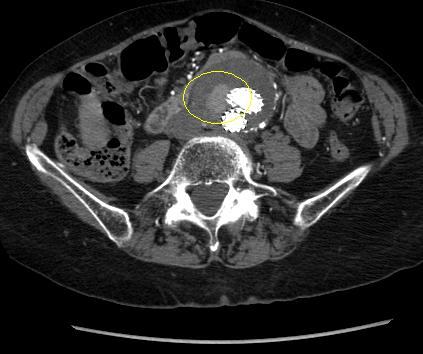
However, most patients with rupture will not survive to reach the operating suite.[Figure caption and citation for the preceding image starts]: Computed tomography scan of a ruptured AAAUniversity of Michigan, specifically the cases of Dr G.R. Upchurch reflecting the Departments of Vascular Surgery and Radiology [Citation ends].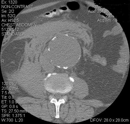
The American College of Cardiology/American Heart Association (ACC/AHA) recommend computed tomography (CT) imaging in patients presenting with ruptured AAA who are hemodynamically stable to evaluate whether the AAA is amenable to endovascular repair.[5]Isselbacher EM, Preventza O, Hamilton Black J 3rd, et al. 2022 ACC/AHA guideline for the diagnosis and management of aortic disease: a report of the American Heart Association/American College of Cardiology Joint Committee on Clinical Practice Guidelines. Circulation. 2022 Dec 13;146(24):e334-e482.
https://www.doi.org/10.1161/CIR.0000000000001106
http://www.ncbi.nlm.nih.gov/pubmed/36322642?tool=bestpractice.com
This recommendation is supported by results from the IMPROVE multicenter randomized controlled trial, which suggest that for most patients, confirmatory CT did not add significant delays to treatment and facilitated appropriate preoperative planning.[108]Powell JT, Hinchcliffe RJ, Thompson MM, et al; IMPROVE Trial Investigators. Observations from the IMPROVE trial concerning the clinical care of patients with ruptured abdominal aortic aneurysm. Br J Surg. 2014 Feb;101(3):216-24.
https://bjssjournals.onlinelibrary.wiley.com/doi/full/10.1002/bjs.9410
http://www.ncbi.nlm.nih.gov/pubmed/24469620?tool=bestpractice.com
If the anatomy is suitable, the ACC/AHA recommend endovascular repair over open repair to reduce the risk of morbidity and mortality.[5]Isselbacher EM, Preventza O, Hamilton Black J 3rd, et al. 2022 ACC/AHA guideline for the diagnosis and management of aortic disease: a report of the American Heart Association/American College of Cardiology Joint Committee on Clinical Practice Guidelines. Circulation. 2022 Dec 13;146(24):e334-e482.
https://www.doi.org/10.1161/CIR.0000000000001106
http://www.ncbi.nlm.nih.gov/pubmed/36322642?tool=bestpractice.com
In patients with confirmed ruptured AAA, 3-year mortality was lower among those randomized to endovascular aneurysm repair (EVAR) than to an open repair strategy (48% vs. 56%; hazard ratio [HR] 0.57, 95% CI 0.36 to 0.90).[109]IMPROVE Trial Investigators. Comparative clinical effectiveness and cost effectiveness of endovascular strategy v open repair for ruptured abdominal aortic aneurysm: three year results of the IMPROVE randomised trial. BMJ. 2017 Nov 14;359:j4859.
https://www.bmj.com/content/359/bmj.j4859.long
http://www.ncbi.nlm.nih.gov/pubmed/29138135?tool=bestpractice.com
The difference between treatment groups was no longer evident after 7 years of follow-up (HR 0.92, 95% CI 0.75 to 1.13). Re-intervention rates were not significantly different between the randomized groups at 3 years (HR 1.02, 95% CI 0.79 to 1.32).[109]IMPROVE Trial Investigators. Comparative clinical effectiveness and cost effectiveness of endovascular strategy v open repair for ruptured abdominal aortic aneurysm: three year results of the IMPROVE randomised trial. BMJ. 2017 Nov 14;359:j4859.
https://www.bmj.com/content/359/bmj.j4859.long
http://www.ncbi.nlm.nih.gov/pubmed/29138135?tool=bestpractice.com
There is some evidence to suggest that an endovascular strategy for repair of ruptured AAA may reduce mortality more effectively in women than in men.[109]IMPROVE Trial Investigators. Comparative clinical effectiveness and cost effectiveness of endovascular strategy v open repair for ruptured abdominal aortic aneurysm: three year results of the IMPROVE randomised trial. BMJ. 2017 Nov 14;359:j4859.
https://www.bmj.com/content/359/bmj.j4859.long
http://www.ncbi.nlm.nih.gov/pubmed/29138135?tool=bestpractice.com
[110]Sweeting MJ, Balm R, Desgranges P, et al; Ruptured Aneurysm Trialists. Individual-patient meta-analysis of three randomized trials comparing endovascular versus open repair for ruptured abdominal aortic aneurysm. Br J Surg. 2015 Sep;102(10):1229-39.
https://bjssjournals.onlinelibrary.wiley.com/doi/full/10.1002/bjs.9852
http://www.ncbi.nlm.nih.gov/pubmed/26104471?tool=bestpractice.com
There is some evidence to suggest that mode of anesthesia for operative repair of AAA affects outcomes.[5]Isselbacher EM, Preventza O, Hamilton Black J 3rd, et al. 2022 ACC/AHA guideline for the diagnosis and management of aortic disease: a report of the American Heart Association/American College of Cardiology Joint Committee on Clinical Practice Guidelines. Circulation. 2022 Dec 13;146(24):e334-e482.
https://www.doi.org/10.1161/CIR.0000000000001106
http://www.ncbi.nlm.nih.gov/pubmed/36322642?tool=bestpractice.com
[111]Armstrong RA, Squire YG, Rogers CA, et al. Type of anesthesia for endovascular abdominal aortic aneurysm repair. J Cardiothorac Vasc Anesth. 2019 Feb;33(2):462-71.
http://www.ncbi.nlm.nih.gov/pubmed/30342821?tool=bestpractice.com
In 2024, the European Society for Vascular Surgery (ESVS) issued a weak recommendation favoring local anesthesia over general anesthesia in elective settings, based on potential reduction in procedure time, ICU admissions, and postoperative hospital stay.[3]Wanhainen A, Van Herzeele I, Bastos Goncalves F, et al. Editor's choice -- European Society for Vascular Surgery (ESVS) 2024 clinical practice guidelines on the management of abdominal aorto-iliac artery aneurysms. Eur J Vasc Endovasc Surg. 2024 Feb;67(2):192-331.
https://www.ejves.com/article/S1078-5884(23)00889-4/fulltext
http://www.ncbi.nlm.nih.gov/pubmed/38307694?tool=bestpractice.com
[112]Liu Y, Wang T, Zhao J, et al. Influence of anesthetic techniques on perioperative outcomes after endovascular aneurysm repair. Ann Vasc Surg. 2021 May;73:375-84.
http://www.ncbi.nlm.nih.gov/pubmed/33383135?tool=bestpractice.com
[113]Zottola ZR, Kruger JL, Kong DS, et al. Locoregional anesthesia is associated with reduced hospital stay and need for intensive care unit care of elective endovascular aneurysm repair patients in the Vascular Quality Initiative. J Vasc Surg. 2023 Apr;77(4):1061-9.
https://www.jvascsurg.org/article/S0741-5214(22)02534-4/fulltext
http://www.ncbi.nlm.nih.gov/pubmed/36400363?tool=bestpractice.com
[114]Kothandan H, Haw Chieh GL, Khan SA, et al. Anesthetic considerations for endovascular abdominal aortic aneurysm repair. Ann Card Anaesth. 2016 Jan-Mar;19(1):132-41.
https://pmc.ncbi.nlm.nih.gov/articles/PMC4900395
http://www.ncbi.nlm.nih.gov/pubmed/26750684?tool=bestpractice.com
The IMPROVE (Immediate Management of Patients with Rupture: Open Versus Endovascular Repair) multicenter randomized controlled trial detected a significantly reduced 30-day mortality in patients who had EVAR under local anesthesia alone compared with general anesthesia (adjusted OR 0.27, 0.1 to 0.7).[108]Powell JT, Hinchcliffe RJ, Thompson MM, et al; IMPROVE Trial Investigators. Observations from the IMPROVE trial concerning the clinical care of patients with ruptured abdominal aortic aneurysm. Br J Surg. 2014 Feb;101(3):216-24.
https://bjssjournals.onlinelibrary.wiley.com/doi/full/10.1002/bjs.9410
http://www.ncbi.nlm.nih.gov/pubmed/24469620?tool=bestpractice.com
A separate meta-analysis comparing mode of anesthesia for endovascular repair of ruptured AAA replicated these findings or improved outcomes with EVAR under local anesthesia.[115]Harky A, Ahmad MU, Santoro G, et al. Local versus general anesthesia in nonemergency endovascular abdominal aortic aneurysm repair: a systematic review and meta-analysis. J Cardiothorac Vasc Anesth. 2020 Apr;34(4):1051-9.
http://www.ncbi.nlm.nih.gov/pubmed/31473112?tool=bestpractice.com
However, another systematic review did not show any mortality benefit with local anesthesia, but did demonstrate shorter hospital stays.[116]Deng J, Liu J, Rong D, et al. A meta-analysis of locoregional anesthesia versus general anesthesia in endovascular repair of ruptured abdominal aortic aneurysm. J Vasc Surg. 2021 Feb;73(2):700-10.
https://www.doi.org/10.1016/j.jvs.2020.08.112
http://www.ncbi.nlm.nih.gov/pubmed/32882348?tool=bestpractice.com
Data from the UK’s National Vascular Registry (9783 patients who received an elective, standard infrarenal EVAR; general anesthetic, n = 7069; regional anesthetic, n = 2347; local anesthetic, n = 367) showed a lower 30 day mortality rate after regional versus general anesthesia.[117]Dovell G, Rogers CA, Armstrong R, et al. The effect of mode of anaesthesia on outcomes after elective endovascular repair of abdominal aortic aneurysm. Eur J Vasc Endovasc Surg. 2020 May;59(5):729-38.
https://www.ejves.com/article/S1078-5884(20)30118-0/fulltext
http://www.ncbi.nlm.nih.gov/pubmed/32291124?tool=bestpractice.com
The international multicenter Endurant Stent Graft Natural Selection Global Post-Market Registry (ENGAGE) study examined the outcomes of 1231 patients undergoing EVAR under general (62% of patients), regional (27%), and local (11%) anesthesia.[118]Broos PP, Stokmans RA, Cuypers PW, et al. Effects of anesthesia type on perioperative outcome after endovascular aneurysm repair. J Endovasc Ther. 2015 Oct;22(5):770-7.
http://www.ncbi.nlm.nih.gov/pubmed/26276553?tool=bestpractice.com
The type of anesthesia had no influence on perioperative mortality or morbidity but the use of local or regional anesthesia during EVAR appeared to be beneficial in decreasing procedure time, need for ICU admission, and duration of postoperative hospital stay.[118]Broos PP, Stokmans RA, Cuypers PW, et al. Effects of anesthesia type on perioperative outcome after endovascular aneurysm repair. J Endovasc Ther. 2015 Oct;22(5):770-7.
http://www.ncbi.nlm.nih.gov/pubmed/26276553?tool=bestpractice.com
Supportive treatment of ruptured AAA
Standard resuscitation measures should be initiated immediately. These include:
Airway management (supplemental oxygen or endotracheal intubation and assisted ventilation if the patient is unconscious).
Intravenous access (central venous catheter).
Arterial catheter; urinary catheter.
Hypotensive resuscitation: aggressive fluid replacement may cause dilutional and hypothermic coagulopathy and secondary clot disruption from increased blood flow, increased perfusion pressure, and decreased blood viscosity, thereby exacerbating bleeding.[119]Roberts K, Revell M, Youssef H, et al. Hypotensive resuscitation in patients with ruptured abdominal aortic aneurysm. Eur J Vasc Endovasc Surg. 2006 Apr;31(4):339-44.
http://www.ncbi.nlm.nih.gov/pubmed/16388972?tool=bestpractice.com
[120]Ohki T, Veith FJ. Endovascular grafts and other image-guided catheter-based adjuncts to improve the treatment of ruptured aortoiliac aneurysms. Ann Surg. 2000 Oct;232(4):466-79.
http://www.ncbi.nlm.nih.gov/pubmed/10998645?tool=bestpractice.com
A target systolic blood pressure (SBP) of 50 to 70 mmHg and withholding fluids is advocated preoperatively.[119]Roberts K, Revell M, Youssef H, et al. Hypotensive resuscitation in patients with ruptured abdominal aortic aneurysm. Eur J Vasc Endovasc Surg. 2006 Apr;31(4):339-44.
http://www.ncbi.nlm.nih.gov/pubmed/16388972?tool=bestpractice.com
[120]Ohki T, Veith FJ. Endovascular grafts and other image-guided catheter-based adjuncts to improve the treatment of ruptured aortoiliac aneurysms. Ann Surg. 2000 Oct;232(4):466-79.
http://www.ncbi.nlm.nih.gov/pubmed/10998645?tool=bestpractice.com
The ACC/AHA guidelines recommend permissive hypotension to reduce bleeding.[5]Isselbacher EM, Preventza O, Hamilton Black J 3rd, et al. 2022 ACC/AHA guideline for the diagnosis and management of aortic disease: a report of the American Heart Association/American College of Cardiology Joint Committee on Clinical Practice Guidelines. Circulation. 2022 Dec 13;146(24):e334-e482.
https://www.doi.org/10.1161/CIR.0000000000001106
http://www.ncbi.nlm.nih.gov/pubmed/36322642?tool=bestpractice.com
However, recommended targets vary and there is no consensus among global guideline groups.
The IMPROVE multicenter randomized controlled trial found that lowest SBP was significantly and independently associated with 30-day mortality in patients with ruptured AAA, and that a target SBP below 70 mmHg in older patients may have contributed to worse outcomes.[108]Powell JT, Hinchcliffe RJ, Thompson MM, et al; IMPROVE Trial Investigators. Observations from the IMPROVE trial concerning the clinical care of patients with ruptured abdominal aortic aneurysm. Br J Surg. 2014 Feb;101(3):216-24.
https://bjssjournals.onlinelibrary.wiley.com/doi/full/10.1002/bjs.9410
http://www.ncbi.nlm.nih.gov/pubmed/24469620?tool=bestpractice.com
In people with ruptured AAA with a recorded preoperative SBP less than 70 mmHg, mortality at 30 days was higher (51.0%) compared with those with SBP above 70 mmHg (34.1%).[108]Powell JT, Hinchcliffe RJ, Thompson MM, et al; IMPROVE Trial Investigators. Observations from the IMPROVE trial concerning the clinical care of patients with ruptured abdominal aortic aneurysm. Br J Surg. 2014 Feb;101(3):216-24.
https://bjssjournals.onlinelibrary.wiley.com/doi/full/10.1002/bjs.9410
http://www.ncbi.nlm.nih.gov/pubmed/24469620?tool=bestpractice.com
One Cochrane review of controlled (permissive) hypotension versus normotensive resuscitative strategy for people with ruptured AAA, which included the IMPROVE trial, noted that people with ruptured AAA are usually older and more likely to have coronary and renal atherosclerotic disease. These patients are also at greater risk of myocardial infarction and renal insufficiency if submitted to low SBP levels compared with younger people with trauma.[121]Moreno DH, Cacione DG, Baptista-Silva JC. Controlled hypotension versus normotensive resuscitation strategy for people with ruptured abdominal aortic aneurysm. Cochrane Database Syst Rev. 2018 Jun 13;6(6):CD011664.
https://www.doi.org/10.1002/14651858.CD011664.pub3
http://www.ncbi.nlm.nih.gov/pubmed/29897100?tool=bestpractice.com
Blood product (packed red cells, platelets, and fresh frozen plasma) availability and transfusion for resuscitation, severe anemia, and coagulopathy.
Notifying anesthetic, intensive care unit (ICU), and operating teams.
Symptomatic but not ruptured AAA
In patients with symptomatic aortic aneurysm, urgent repair is indicated regardless of diameter.[5]Isselbacher EM, Preventza O, Hamilton Black J 3rd, et al. 2022 ACC/AHA guideline for the diagnosis and management of aortic disease: a report of the American Heart Association/American College of Cardiology Joint Committee on Clinical Practice Guidelines. Circulation. 2022 Dec 13;146(24):e334-e482.
https://www.doi.org/10.1161/CIR.0000000000001106
http://www.ncbi.nlm.nih.gov/pubmed/36322642?tool=bestpractice.com
[78]National Institute for Health and Care Excellence. Abdominal aortic aneurysm: diagnosis and management. Mar 2020 [internet publication].
https://www.nice.org.uk/guidance/ng156
[102]Mazzolai L, Teixido-Tura G, Lanzi S, et al. 2024 ESC guidelines for the management of peripheral arterial and aortic diseases. Eur Heart J. 2024 Sep 29;45(36):3538-700.
https://academic.oup.com/eurheartj/article/45/36/3538/7738955
The development of new or worsening pain may herald aneurysm expansion and impending rupture. Symptomatic, nonruptured aneurysm is, therefore, best treated urgently.[76]Chaikof EL, Dalman RL, Eskandari MK, et al. The Society for Vascular Surgery practice guidelines on the care of patients with an abdominal aortic aneurysm. J Vasc Surg. 2018 Jan;67(1):2-77.e2.
https://www.jvascsurg.org/article/S0741-5214(17)32369-8/fulltext
http://www.ncbi.nlm.nih.gov/pubmed/29268916?tool=bestpractice.com
Under some circumstances, intervention may be delayed for several hours to optimize conditions to ensure successful repair; these patients should be closely monitored in the ICU.[76]Chaikof EL, Dalman RL, Eskandari MK, et al. The Society for Vascular Surgery practice guidelines on the care of patients with an abdominal aortic aneurysm. J Vasc Surg. 2018 Jan;67(1):2-77.e2.
https://www.jvascsurg.org/article/S0741-5214(17)32369-8/fulltext
http://www.ncbi.nlm.nih.gov/pubmed/29268916?tool=bestpractice.com
EVAR is increasingly used in the management of patients with symptomatic AAA.[125]De Martino RR, Nolan BW, Goodney PP, Chang CK, et al; Vascular Study Group of Northern New England. Outcomes of symptomatic abdominal aortic aneurysm repair. J Vasc Surg. 2010 Jul;52(1):5-12.e1.
https://www.jvascsurg.org/article/S0741-5214(10)00259-4/fulltext
http://www.ncbi.nlm.nih.gov/pubmed/20471771?tool=bestpractice.com
[126]Chandra V, Trang K, Virgin-Downey W, et al. Management and outcomes of symptomatic abdominal aortic aneurysms during the past 20 years. J Vasc Surg. 2017 Dec;66(6):1679-85.
http://www.ncbi.nlm.nih.gov/pubmed/28619644?tool=bestpractice.com
In observational studies, short-term all-cause mortality rates did not differ between endovascular and open repair of symptomatic AAA.[125]De Martino RR, Nolan BW, Goodney PP, Chang CK, et al; Vascular Study Group of Northern New England. Outcomes of symptomatic abdominal aortic aneurysm repair. J Vasc Surg. 2010 Jul;52(1):5-12.e1.
https://www.jvascsurg.org/article/S0741-5214(10)00259-4/fulltext
http://www.ncbi.nlm.nih.gov/pubmed/20471771?tool=bestpractice.com
[126]Chandra V, Trang K, Virgin-Downey W, et al. Management and outcomes of symptomatic abdominal aortic aneurysms during the past 20 years. J Vasc Surg. 2017 Dec;66(6):1679-85.
http://www.ncbi.nlm.nih.gov/pubmed/28619644?tool=bestpractice.com
[127]Ten Bosch JA, Willigendael EM, Kruidenier LM, et al. Early and mid-term results of a prospective observational study comparing emergency endovascular aneurysm repair with open surgery in both ruptured and unruptured acute abdominal aortic aneurysms. Vascular. 2012 Apr;20(2):72-80.
http://www.ncbi.nlm.nih.gov/pubmed/22454547?tool=bestpractice.com
Data from the 2011-2013 American College of Surgeons National Surgical Quality Improvement Program suggest that 30-day mortality risk after repair of symptomatic AAA was approximately double that following asymptomatic AAA repair, regardless of surgical approach (EVAR: symptomatic 3.8% vs. asymptomatic 1.4%, P=0.001; open surgery: symptomatic 7.7% vs. asymptomatic 4.3%, P=0.08).[128]Soden PA, Zettervall SL, Ultee KH, et al. Outcomes for symptomatic abdominal aortic aneurysms in the American College of Surgeons National Surgical Quality Improvement Program. J Vasc Surg. 2016 Aug;64(2):297-305.
https://www.jvascsurg.org/article/S0741-5214(16)00320-7/fulltext
http://www.ncbi.nlm.nih.gov/pubmed/27146791?tool=bestpractice.com
Smaller patient numbers likely contribute to the non-statistically significant finding reported for open repair.
Incidental finding of small asymptomatic AAA
For AAA detected as an incidental finding, surveillance is preferred to repair until the theoretical risk of rupture exceeds the estimated risk of operative mortality.[4]Owens DK, Davidson KW, Krist AH, et al; US Preventive Services Task Force. Screening for abdominal aortic aneurysm: US Preventive Services Task Force recommendation statement. JAMA. 2019 Dec 10;322(22):2211-8.
https://jamanetwork.com/journals/jama/fullarticle/2757234
http://www.ncbi.nlm.nih.gov/pubmed/31821437?tool=bestpractice.com
Early surgery for the treatment of smaller AAAs does not reduce all-cause or AAA-specific mortality.[4]Owens DK, Davidson KW, Krist AH, et al; US Preventive Services Task Force. Screening for abdominal aortic aneurysm: US Preventive Services Task Force recommendation statement. JAMA. 2019 Dec 10;322(22):2211-8.
https://jamanetwork.com/journals/jama/fullarticle/2757234
http://www.ncbi.nlm.nih.gov/pubmed/31821437?tool=bestpractice.com
[129]Ulug P, Powell JT, Martinez MA, et al. Surgery for small asymptomatic abdominal aortic aneurysms. Cochrane Database Syst Rev. 2020 Jul 1;7(7):CD001835.
https://www.doi.org/10.1002/14651858.CD001835.pub5
http://www.ncbi.nlm.nih.gov/pubmed/32609382?tool=bestpractice.com
[  ]
How does immediate surgery compare with surveillance in people with asymptomatic abdominal aortic aneurysms (AAAs)?/cca.html?targetUrl=https://www.cochranelibrary.com/cca/doi/10.1002/cca.3227/fullShow me the answer One systematic review (4 trials, 3314 participants) found high-quality evidence to demonstrate that immediate repair of small AAA (4 cm to 5.5 cm) did not improve long-term survival compared with surveillance (adjusted HR 0.88, 95% CI 0.75 to 1.02, mean follow-up 10 years).[129]Ulug P, Powell JT, Martinez MA, et al. Surgery for small asymptomatic abdominal aortic aneurysms. Cochrane Database Syst Rev. 2020 Jul 1;7(7):CD001835.
https://www.doi.org/10.1002/14651858.CD001835.pub5
http://www.ncbi.nlm.nih.gov/pubmed/32609382?tool=bestpractice.com
The lack of benefit attributable to immediate surgery was consistent regardless of patient age, diameter of small aneurysm, and whether repair was endovascular or open.[129]Ulug P, Powell JT, Martinez MA, et al. Surgery for small asymptomatic abdominal aortic aneurysms. Cochrane Database Syst Rev. 2020 Jul 1;7(7):CD001835.
https://www.doi.org/10.1002/14651858.CD001835.pub5
http://www.ncbi.nlm.nih.gov/pubmed/32609382?tool=bestpractice.com
]
How does immediate surgery compare with surveillance in people with asymptomatic abdominal aortic aneurysms (AAAs)?/cca.html?targetUrl=https://www.cochranelibrary.com/cca/doi/10.1002/cca.3227/fullShow me the answer One systematic review (4 trials, 3314 participants) found high-quality evidence to demonstrate that immediate repair of small AAA (4 cm to 5.5 cm) did not improve long-term survival compared with surveillance (adjusted HR 0.88, 95% CI 0.75 to 1.02, mean follow-up 10 years).[129]Ulug P, Powell JT, Martinez MA, et al. Surgery for small asymptomatic abdominal aortic aneurysms. Cochrane Database Syst Rev. 2020 Jul 1;7(7):CD001835.
https://www.doi.org/10.1002/14651858.CD001835.pub5
http://www.ncbi.nlm.nih.gov/pubmed/32609382?tool=bestpractice.com
The lack of benefit attributable to immediate surgery was consistent regardless of patient age, diameter of small aneurysm, and whether repair was endovascular or open.[129]Ulug P, Powell JT, Martinez MA, et al. Surgery for small asymptomatic abdominal aortic aneurysms. Cochrane Database Syst Rev. 2020 Jul 1;7(7):CD001835.
https://www.doi.org/10.1002/14651858.CD001835.pub5
http://www.ncbi.nlm.nih.gov/pubmed/32609382?tool=bestpractice.com
Surgical referral of smaller AAA is usually reserved for rapid growth, or once the threshold diameter for aneurysm repair is reached on repeated ultrasonography.[4]Owens DK, Davidson KW, Krist AH, et al; US Preventive Services Task Force. Screening for abdominal aortic aneurysm: US Preventive Services Task Force recommendation statement. JAMA. 2019 Dec 10;322(22):2211-8.
https://jamanetwork.com/journals/jama/fullarticle/2757234
http://www.ncbi.nlm.nih.gov/pubmed/31821437?tool=bestpractice.com
However, in patients with an underlying genetic cause or connective tissue disorder, the threshold diameter for considering repair should be individualized, depending on:[3]Wanhainen A, Van Herzeele I, Bastos Goncalves F, et al. Editor's choice -- European Society for Vascular Surgery (ESVS) 2024 clinical practice guidelines on the management of abdominal aorto-iliac artery aneurysms. Eur J Vasc Endovasc Surg. 2024 Feb;67(2):192-331.
https://www.ejves.com/article/S1078-5884(23)00889-4/fulltext
http://www.ncbi.nlm.nih.gov/pubmed/38307694?tool=bestpractice.com
Anatomic features
Underlying genetics: rupture risk is higher at smaller aortic diameters in some conditions, and surgical repair is more challenging in certain disorders owing to the increased arterial wall fragility and anatomy[3]Wanhainen A, Van Herzeele I, Bastos Goncalves F, et al. Editor's choice -- European Society for Vascular Surgery (ESVS) 2024 clinical practice guidelines on the management of abdominal aorto-iliac artery aneurysms. Eur J Vasc Endovasc Surg. 2024 Feb;67(2):192-331.
https://www.ejves.com/article/S1078-5884(23)00889-4/fulltext
http://www.ncbi.nlm.nih.gov/pubmed/38307694?tool=bestpractice.com
Medical goals for asymptomatic small aneurysms include:
1. Surveillance:
American College of Cardiology Foundation/American Heart Association guidelines recommend that infra-/juxtarenal AAAs measuring 4.0 to 4.9 cm in diameter by ultrasound/CT should be monitored every 6-12 months.[5]Isselbacher EM, Preventza O, Hamilton Black J 3rd, et al. 2022 ACC/AHA guideline for the diagnosis and management of aortic disease: a report of the American Heart Association/American College of Cardiology Joint Committee on Clinical Practice Guidelines. Circulation. 2022 Dec 13;146(24):e334-e482.
https://www.doi.org/10.1161/CIR.0000000000001106
http://www.ncbi.nlm.nih.gov/pubmed/36322642?tool=bestpractice.com
Once larger than 4.5 cm in women and 5 cm in men, these guidelines recommend surveillance every 6 months.[5]Isselbacher EM, Preventza O, Hamilton Black J 3rd, et al. 2022 ACC/AHA guideline for the diagnosis and management of aortic disease: a report of the American Heart Association/American College of Cardiology Joint Committee on Clinical Practice Guidelines. Circulation. 2022 Dec 13;146(24):e334-e482.
https://www.doi.org/10.1161/CIR.0000000000001106
http://www.ncbi.nlm.nih.gov/pubmed/36322642?tool=bestpractice.com
AAAs <3.9 cm can be monitored with ultrasound every 2-3 years.[5]Isselbacher EM, Preventza O, Hamilton Black J 3rd, et al. 2022 ACC/AHA guideline for the diagnosis and management of aortic disease: a report of the American Heart Association/American College of Cardiology Joint Committee on Clinical Practice Guidelines. Circulation. 2022 Dec 13;146(24):e334-e482.
https://www.doi.org/10.1161/CIR.0000000000001106
http://www.ncbi.nlm.nih.gov/pubmed/36322642?tool=bestpractice.com
The UK National Health Service recommends that annual screening intervals are employed for 3.0 to 4.4 cm AAAs and 3-month intervals for 4.5 to 5.4 cm AAAs.[130]Public Health England. NHS public health functions agreement 2019-20. Service specification no.23. NHS Abdominal Aortic Aneurysm Screening Programme. July 2019 [internet publication].
https://www.england.nhs.uk/wp-content/uploads/2017/04/Service-Specification-No.23-Abdominal_Aortic_Aneurysm.pdf
One systematic review and meta-analysis of individual patient data concluded that surveillance intervals of 2 years for 3.0 to 4.4 cm AAA, and 6 months for 4.5 to 5.4 cm AAA, are safe and cost-effective.[131]Thompson S, Brown L, Sweeting M, et al; RESCAN Collaborators. Systematic review and meta-analysis of the growth and rupture rates of small abdominal aortic aneurysms: implications for surveillance intervals and their cost-effectiveness. Health Technol Assess. 2013 Sep;17(41):1-118.
https://www.journalslibrary.nihr.ac.uk/hta/hta17410/#/full-report
http://www.ncbi.nlm.nih.gov/pubmed/24067626?tool=bestpractice.com
Analysis of AAA growth and rupture rates indicated that in order to maintain a AAA rupture risk <1%, an 8.5-year surveillance interval is required for men with baseline AAA diameter of 3.0 cm.[131]Thompson S, Brown L, Sweeting M, et al; RESCAN Collaborators. Systematic review and meta-analysis of the growth and rupture rates of small abdominal aortic aneurysms: implications for surveillance intervals and their cost-effectiveness. Health Technol Assess. 2013 Sep;17(41):1-118.
https://www.journalslibrary.nihr.ac.uk/hta/hta17410/#/full-report
http://www.ncbi.nlm.nih.gov/pubmed/24067626?tool=bestpractice.com
The corresponding estimated surveillance interval for men with an initial aneurysm diameter of 5.0 cm was 17 months. Despite having similar small aneurysm growth rates, rupture rates were four times higher in women than in men.[131]Thompson S, Brown L, Sweeting M, et al; RESCAN Collaborators. Systematic review and meta-analysis of the growth and rupture rates of small abdominal aortic aneurysms: implications for surveillance intervals and their cost-effectiveness. Health Technol Assess. 2013 Sep;17(41):1-118.
https://www.journalslibrary.nihr.ac.uk/hta/hta17410/#/full-report
http://www.ncbi.nlm.nih.gov/pubmed/24067626?tool=bestpractice.com
Surveillance programs and criteria for considering surgery need to be tailored for women with opportunistically detected AAA.
Most AAAs show linear growth; modeling based on this suggests that smaller AAAs (<4.25 cm) could be followed up every 2 years, with minimal chance of exceeding interventional thresholds within that time.[132]Kim GY, Corriere MA. Balancing watching vs waiting during imaging surveillance of small abdominal aortic aneurysms. JAMA Surg. 2021 Apr 1;156(4):370-1.
http://www.ncbi.nlm.nih.gov/pubmed/33595617?tool=bestpractice.com
[133]Olson SL, Wijesinha MA, Panthofer AM, et al. Evaluating growth patterns of abdominal aortic aneurysm diameter with serial computed tomography surveillance. JAMA Surg. 2021 Apr 1;156(4):363-70.
https://www.doi.org/10.1001/jamasurg.2020.7190
http://www.ncbi.nlm.nih.gov/pubmed/33595625?tool=bestpractice.com
Aneurysmal growth of ≥0.5 cm in 6 months may be an indication for repair, to reduce the risk of rupture.[5]Isselbacher EM, Preventza O, Hamilton Black J 3rd, et al. 2022 ACC/AHA guideline for the diagnosis and management of aortic disease: a report of the American Heart Association/American College of Cardiology Joint Committee on Clinical Practice Guidelines. Circulation. 2022 Dec 13;146(24):e334-e482.
https://www.doi.org/10.1161/CIR.0000000000001106
http://www.ncbi.nlm.nih.gov/pubmed/36322642?tool=bestpractice.com
The ESVS recommends incorporation of subaneurysmal aortas (2.5 to 2.9 cm) into AAA surveillance recommendations since long-term cohort studies show that most subaneurysmal aortas eventually progress to an AAA of which a substantial proportion will reach the diameter threshold for consideration of repair.[3]Wanhainen A, Van Herzeele I, Bastos Goncalves F, et al. Editor's choice -- European Society for Vascular Surgery (ESVS) 2024 clinical practice guidelines on the management of abdominal aorto-iliac artery aneurysms. Eur J Vasc Endovasc Surg. 2024 Feb;67(2):192-331.
https://www.ejves.com/article/S1078-5884(23)00889-4/fulltext
http://www.ncbi.nlm.nih.gov/pubmed/38307694?tool=bestpractice.com
Surveillance decisions should take into account life expectancy, suitability for future repair, and patient preferences.[3]Wanhainen A, Van Herzeele I, Bastos Goncalves F, et al. Editor's choice -- European Society for Vascular Surgery (ESVS) 2024 clinical practice guidelines on the management of abdominal aorto-iliac artery aneurysms. Eur J Vasc Endovasc Surg. 2024 Feb;67(2):192-331.
https://www.ejves.com/article/S1078-5884(23)00889-4/fulltext
http://www.ncbi.nlm.nih.gov/pubmed/38307694?tool=bestpractice.com
2. Control of modifiable risk factors for expansion and rupture:
Smoking cessation - nicotine-replacement therapy, nortriptyline, and bupropion, or counseling.[1]Dehlin JM, Upchurch GR. Management of abdominal aortic aneurysms. Curr Treat Options Cardiovasc Med. 2005 Jun;7(2):119-30.
http://www.ncbi.nlm.nih.gov/pubmed/15935120?tool=bestpractice.com
[5]Isselbacher EM, Preventza O, Hamilton Black J 3rd, et al. 2022 ACC/AHA guideline for the diagnosis and management of aortic disease: a report of the American Heart Association/American College of Cardiology Joint Committee on Clinical Practice Guidelines. Circulation. 2022 Dec 13;146(24):e334-e482.
https://www.doi.org/10.1161/CIR.0000000000001106
http://www.ncbi.nlm.nih.gov/pubmed/36322642?tool=bestpractice.com
[13]Zankl AR, Schumacher H, Krumsdorf U, et al. Pathology, natural history and treatment of abdominal aortic aneurysms. Clin Res Cardiol. 2007 Mar;96(3):140-51.
http://www.ncbi.nlm.nih.gov/pubmed/17180573?tool=bestpractice.com
[15]Singh K, Bønaa H, Jacobsen BK, et al. Prevalence of and risk factors for abdominal aortic aneurysms in a population-based study: the Tromsø Study. Am J Epidemiol. 2001 Aug 1;154(3):236-44.
https://academic.oup.com/aje/article/154/3/236/125840
http://www.ncbi.nlm.nih.gov/pubmed/11479188?tool=bestpractice.com
[22]Lederle FA, Johnson GR, Wilson SE, et al; Aneurysm Detection and Management (ADAM) Veterans Affairs Cooperative Study Group. Prevalence and associations of abdominal aortic aneurysm detected through screening. Ann Intern Med. 1997 Mar 15;126(6):441-9.
http://www.ncbi.nlm.nih.gov/pubmed/9072929?tool=bestpractice.com
[23]Wilmink TB, Quick CR, Day NE. The association between cigarette smoking and abdominal aortic aneurysms. J Vasc Surg. 1999 Dec;30(6):1099-105.
http://www.ncbi.nlm.nih.gov/pubmed/10587395?tool=bestpractice.com
[134]Hartmann-Boyce J, Chepkin SC, Ye W, et al. Nicotine replacement therapy versus control for smoking cessation. Cochrane Database Syst Rev. 2018 May 31;5:CD000146.
https://www.cochranelibrary.com/cdsr/doi/10.1002/14651858.CD000146.pub5/full
http://www.ncbi.nlm.nih.gov/pubmed/29852054?tool=bestpractice.com
[135]Rigotti NA, Clair C, Munafò MR, et al. Interventions for smoking cessation in hospitalised patients. Cochrane Database Syst Rev. 2012 May 16;(5):CD001837.
https://www.cochranelibrary.com/cdsr/doi/10.1002/14651858.CD001837.pub3/full
http://www.ncbi.nlm.nih.gov/pubmed/22592676?tool=bestpractice.com
[136]Howes S, Hartmann-Boyce J, Livingstone-Banks J, et al. Antidepressants for smoking cessation. Cochrane Database Syst Rev. 2020 Apr 22;(4):CD000031.
https://www.cochranelibrary.com/cdsr/doi/10.1002/14651858.CD000031.pub5/full
http://www.ncbi.nlm.nih.gov/pubmed/32319681?tool=bestpractice.com
[ ]
What are the effects of adding bupropion or fluoxetine to other treatments compared with using other treatments alone for people trying to quit smoking?/cca.html?targetUrl=https://www.cochranelibrary.com/cca/doi/10.1002/cca.4337/fullShow me the answer
Short-term treatment with beta-blockers does not appear to reduce the rate of AAA expansion.[4]Owens DK, Davidson KW, Krist AH, et al; US Preventive Services Task Force. Screening for abdominal aortic aneurysm: US Preventive Services Task Force recommendation statement. JAMA. 2019 Dec 10;322(22):2211-8.
https://jamanetwork.com/journals/jama/fullarticle/2757234
http://www.ncbi.nlm.nih.gov/pubmed/31821437?tool=bestpractice.com
[137]Siordia JA. Beta-blockers and abdominal aortic aneurysm growth: a systematic review and meta-analysis. Curr Cardiol Rev. 2021;17(4):e230421187502.
http://www.ncbi.nlm.nih.gov/pubmed/33143615?tool=bestpractice.com
Trials in which patients with small AAAs were randomized to propranolol, and other beta-blockers, with the intention of reducing the rate of aneurysm expansion failed to demonstrate significant protective effects.[137]Siordia JA. Beta-blockers and abdominal aortic aneurysm growth: a systematic review and meta-analysis. Curr Cardiol Rev. 2021;17(4):e230421187502.
http://www.ncbi.nlm.nih.gov/pubmed/33143615?tool=bestpractice.com
[138]Rughani G, Robertson L, Clarke M. Medical treatment for small abdominal aortic aneurysms. Cochrane Database Syst Rev. 2012 Sep 12;(9):CD009536.
https://www.cochranelibrary.com/cdsr/doi/10.1002/14651858.CD009536.pub2/full
http://www.ncbi.nlm.nih.gov/pubmed/22972146?tool=bestpractice.com
Propranolol was poorly tolerated in these studies.[138]Rughani G, Robertson L, Clarke M. Medical treatment for small abdominal aortic aneurysms. Cochrane Database Syst Rev. 2012 Sep 12;(9):CD009536.
https://www.cochranelibrary.com/cdsr/doi/10.1002/14651858.CD009536.pub2/full
http://www.ncbi.nlm.nih.gov/pubmed/22972146?tool=bestpractice.com
3. Aggressive management of other cardiovascular disease:
Modifiable cardiovascular risk factors such as hypertension and hyperlipidemia should be treated.[5]Isselbacher EM, Preventza O, Hamilton Black J 3rd, et al. 2022 ACC/AHA guideline for the diagnosis and management of aortic disease: a report of the American Heart Association/American College of Cardiology Joint Committee on Clinical Practice Guidelines. Circulation. 2022 Dec 13;146(24):e334-e482.
https://www.doi.org/10.1161/CIR.0000000000001106
http://www.ncbi.nlm.nih.gov/pubmed/36322642?tool=bestpractice.com
[78]National Institute for Health and Care Excellence. Abdominal aortic aneurysm: diagnosis and management. Mar 2020 [internet publication].
https://www.nice.org.uk/guidance/ng156
Statins should be started at least 1 month before surgery to reduce cardiovascular morbidity and mortality, and continued indefinitely.[3]Wanhainen A, Van Herzeele I, Bastos Goncalves F, et al. Editor's choice -- European Society for Vascular Surgery (ESVS) 2024 clinical practice guidelines on the management of abdominal aorto-iliac artery aneurysms. Eur J Vasc Endovasc Surg. 2024 Feb;67(2):192-331.
https://www.ejves.com/article/S1078-5884(23)00889-4/fulltext
http://www.ncbi.nlm.nih.gov/pubmed/38307694?tool=bestpractice.com
[139]Risum Ø, Sandven I, Sundhagen JO, et al. Editor's choice - effect of statins on total mortality in abdominal aortic aneurysm repair: a systematic review and meta-analysis. Eur J Vasc Endovasc Surg. 2021 Jan;61(1):114-20.
https://www.doi.org/10.1016/j.ejvs.2020.08.007
http://www.ncbi.nlm.nih.gov/pubmed/32928667?tool=bestpractice.com
There is limited evidence, but in the absence of any contraindication, patients with AAA should receive single antiplatelet therapy (aspirin or clopidogrel).[3]Wanhainen A, Van Herzeele I, Bastos Goncalves F, et al. Editor's choice -- European Society for Vascular Surgery (ESVS) 2024 clinical practice guidelines on the management of abdominal aorto-iliac artery aneurysms. Eur J Vasc Endovasc Surg. 2024 Feb;67(2):192-331.
https://www.ejves.com/article/S1078-5884(23)00889-4/fulltext
http://www.ncbi.nlm.nih.gov/pubmed/38307694?tool=bestpractice.com
[5]Isselbacher EM, Preventza O, Hamilton Black J 3rd, et al. 2022 ACC/AHA guideline for the diagnosis and management of aortic disease: a report of the American Heart Association/American College of Cardiology Joint Committee on Clinical Practice Guidelines. Circulation. 2022 Dec 13;146(24):e334-e482.
https://www.doi.org/10.1161/CIR.0000000000001106
http://www.ncbi.nlm.nih.gov/pubmed/36322642?tool=bestpractice.com
[140]Aboyans V, Bauersachs R, Mazzolai L, et al. Antithrombotic therapies in aortic and peripheral arterial diseases in 2021: a consensus document from the ESC working group on aorta and peripheral vascular diseases, the ESC working group on thrombosis, and the ESC working group on cardiovascular pharmacotherapy. Eur Heart J. 2021 Oct 14;42(39):4013-24.
https://www.doi.org/10.1093/eurheartj/ehab390
http://www.ncbi.nlm.nih.gov/pubmed/34279602?tool=bestpractice.com
Incidental finding of large asymptomatic AAA
Generally, repair is indicated in patients with large asymptomatic AAA (e.g., with a diameter exceeding 5.5 cm in men or 5.0 cm in women in the US, although treatment decisions based on greater size may differ in other countries).[5]Isselbacher EM, Preventza O, Hamilton Black J 3rd, et al. 2022 ACC/AHA guideline for the diagnosis and management of aortic disease: a report of the American Heart Association/American College of Cardiology Joint Committee on Clinical Practice Guidelines. Circulation. 2022 Dec 13;146(24):e334-e482.
https://www.doi.org/10.1161/CIR.0000000000001106
http://www.ncbi.nlm.nih.gov/pubmed/36322642?tool=bestpractice.com
Repair of aneurysms ≥5.5 cm offers a survival advantage.[1]Dehlin JM, Upchurch GR. Management of abdominal aortic aneurysms. Curr Treat Options Cardiovasc Med. 2005 Jun;7(2):119-30.
http://www.ncbi.nlm.nih.gov/pubmed/15935120?tool=bestpractice.com
[76]Chaikof EL, Dalman RL, Eskandari MK, et al. The Society for Vascular Surgery practice guidelines on the care of patients with an abdominal aortic aneurysm. J Vasc Surg. 2018 Jan;67(1):2-77.e2.
https://www.jvascsurg.org/article/S0741-5214(17)32369-8/fulltext
http://www.ncbi.nlm.nih.gov/pubmed/29268916?tool=bestpractice.com
[78]National Institute for Health and Care Excellence. Abdominal aortic aneurysm: diagnosis and management. Mar 2020 [internet publication].
https://www.nice.org.uk/guidance/ng156
[104]UK Small Aneurysm Trial Participants. Mortality results for randomized controlled trial of early elective surgery or ultrasonographic surveillance for small abdominal aortic aneurysms. Lancet. 1998 Nov 21;352(9141):1649-55.
http://www.ncbi.nlm.nih.gov/pubmed/9853436?tool=bestpractice.com
[105]Powell JT, Brady AR, Brown LC, et al; United Kingdom Small Aneurysm Trial Participants. Long-term outcomes of immediate repair compared with surveillance of small abdominal aortic aneurysms. N Engl J Med. 2002 May 9;346(19):1445-52.
https://www.nejm.org/doi/full/10.1056/NEJMoa013527
http://www.ncbi.nlm.nih.gov/pubmed/12000814?tool=bestpractice.com
[106]Powell JT, Brown LC, Forbes JF, et al. Final 12-year follow-up of surgery versus surveillance in the UK Small Aneurysm Trial. Br J Surg. 2007 Jun;94(6):702-8.
http://www.ncbi.nlm.nih.gov/pubmed/17514693?tool=bestpractice.com
Decisions regarding repair should be individualized, taking account of patient preference, patient age, sex, perioperative risk factors, and anatomic risk factors. Care should be taken to evaluate patient quality of life, and careful counseling undertaken regarding the risks of surgery (e.g., informing patients of their Vascular Quality Initiative perioperative mortality risk score) and subsequent quality of life. A shared decision making approach taking into account the risks and benefits of the procedures is recommended.[5]Isselbacher EM, Preventza O, Hamilton Black J 3rd, et al. 2022 ACC/AHA guideline for the diagnosis and management of aortic disease: a report of the American Heart Association/American College of Cardiology Joint Committee on Clinical Practice Guidelines. Circulation. 2022 Dec 13;146(24):e334-e482.
https://www.doi.org/10.1161/CIR.0000000000001106
http://www.ncbi.nlm.nih.gov/pubmed/36322642?tool=bestpractice.com
EVAR should be considered in patients who are unfit for open surgery.[5]Isselbacher EM, Preventza O, Hamilton Black J 3rd, et al. 2022 ACC/AHA guideline for the diagnosis and management of aortic disease: a report of the American Heart Association/American College of Cardiology Joint Committee on Clinical Practice Guidelines. Circulation. 2022 Dec 13;146(24):e334-e482.
https://www.doi.org/10.1161/CIR.0000000000001106
http://www.ncbi.nlm.nih.gov/pubmed/36322642?tool=bestpractice.com
[76]Chaikof EL, Dalman RL, Eskandari MK, et al. The Society for Vascular Surgery practice guidelines on the care of patients with an abdominal aortic aneurysm. J Vasc Surg. 2018 Jan;67(1):2-77.e2.
https://www.jvascsurg.org/article/S0741-5214(17)32369-8/fulltext
http://www.ncbi.nlm.nih.gov/pubmed/29268916?tool=bestpractice.com
[78]National Institute for Health and Care Excellence. Abdominal aortic aneurysm: diagnosis and management. Mar 2020 [internet publication].
https://www.nice.org.uk/guidance/ng156
[129]Ulug P, Powell JT, Martinez MA, et al. Surgery for small asymptomatic abdominal aortic aneurysms. Cochrane Database Syst Rev. 2020 Jul 1;7(7):CD001835.
https://www.doi.org/10.1002/14651858.CD001835.pub5
http://www.ncbi.nlm.nih.gov/pubmed/32609382?tool=bestpractice.com
Data suggest that in patients with large AAAs (ranging from 5.0 to 5.5 cm) undergoing elective repair, EVAR is equivalent to open repair in terms of overall survival, although the rate of secondary interventions is higher for EVAR.[141]Greenhalgh RM, Brown LC, Powell JT, et al; United Kingdom EVAR Trial Investigators. Endovascular versus open repair of abdominal aortic aneurysm. N Engl J Med. 2010 May 20;362(20):1863-71.
http://www.ncbi.nlm.nih.gov/pubmed/20382983?tool=bestpractice.com
[142]Amato B, Fugetto F, Compagna R, et al. Endovascular repair versus open repair in the treatment of ruptured aortic aneurysms: a systematic review. Minerva Chir. 2019 Dec;74(6):472-80.
http://www.ncbi.nlm.nih.gov/pubmed/29806754?tool=bestpractice.com
EVAR reduces AAA-related mortality (but not longer-term overall survival) in patients with large AAA (≥5.5 cm) who are unsuitable for open repair.[143]Greenhalgh RM, Brown LC, Powell JT, et al; United Kingdom EVAR Trial Investigators. Endovascular repair of aortic aneurysm in patients physically ineligible for open repair. N Engl J Med. 2010 May 20;362(20):1872-80.
http://www.ncbi.nlm.nih.gov/pubmed/20382982?tool=bestpractice.com
Post-repair, larger AAAs appear to be associated with worse late survival than smaller aneurysms (pooled HR 1.14 per 1-cm increase in AAA diameter, 95% CI 1.09 to 1.18; 12.0- to 91.2-month follow-up).[144]Khashram M, Hider PN, Williman JA, et al. Does the diameter of abdominal aortic aneurysm influence late survival following abdominal aortic aneurysm repair? A systematic review and meta-analysis. Vascular. 2016 Dec;24(6):658-67.
http://www.ncbi.nlm.nih.gov/pubmed/27189809?tool=bestpractice.com
The association is more pronounced with EVAR than with open repair.
Elective repair in asymptomatic patients allows for preoperative assessment, cardiac risk stratification, and medical optimization of other comorbidities. Coronary artery disease remains the leading cause of early and late mortality after AAA repair.
Endovascular aneurysm repair (EVAR)
EVAR involves the transfemoral endoluminal delivery of a covered stent graft into the aorta, thus sealing off the aneurysm wall from systemic pressures, preventing rupture, and allowing for sac shrinkage. The endograft can be deployed percutaneously through low-profile devices, or after exposing the femoral arteries surgically. A Cochrane review found no difference between the techniques after short follow-up (6 months), except that the percutaneous approach may be faster.[145]Gimzewska M, Jackson AI, Yeoh SE, et al. Totally percutaneous versus surgical cut-down femoral artery access for elective bifurcated abdominal endovascular aneurysm repair. Cochrane Database Syst Rev. 2017 Feb 21;(2):CD010185.
https://www.cochranelibrary.com/cdsr/doi/10.1002/14651858.CD010185.pub3/full
http://www.ncbi.nlm.nih.gov/pubmed/28221665?tool=bestpractice.com
[ ]
How does totally percutaneous femoral artery access compare with surgical cut‐down femoral artery access for adults undergoing elective bifurcated abdominal endovascular aneurysm repair?/cca.html?targetUrl=https://www.cochranelibrary.com/cca/doi/10.1002/cca.4261/fullShow me the answer Long-term data for the durability of low-profile devices are lacking.[Figure caption and citation for the preceding image starts]: Various endovascular stent grafts used for endovascular aneurysm repair (EVAR)University of Michigan/Dr G.R. Upchurch, Departments of Vascular Surgery and Radiology [Citation ends].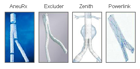 [Figure caption and citation for the preceding image starts]: Endovascular aneurysm repair (EVAR)University of Michigan/Dr G.R. Upchurch, Departments of Vascular Surgery and Radiology [Citation ends].
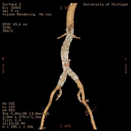
Assessment of suitability for EVAR should be through the use of 0.5 mm-slice CT angiography.[79]American College of Radiology. ACR Aappropriateness Criteria: pulsatile abdominal mass, suspected abdominal aortic aneurysm. 2023 [internet publication].
https://acsearch.acr.org/docs/69414/Narrative
It is essential that the operator is familiar with the specific instructions for use of the endograft to be used.
Lifelong, annual surveillance with ultrasonography or CT is recommended following endovascular repair of AAAs to detect late complications and identify late device failure and disease progression.[3]Wanhainen A, Van Herzeele I, Bastos Goncalves F, et al. Editor's choice -- European Society for Vascular Surgery (ESVS) 2024 clinical practice guidelines on the management of abdominal aorto-iliac artery aneurysms. Eur J Vasc Endovasc Surg. 2024 Feb;67(2):192-331.
https://www.ejves.com/article/S1078-5884(23)00889-4/fulltext
http://www.ncbi.nlm.nih.gov/pubmed/38307694?tool=bestpractice.com
[76]Chaikof EL, Dalman RL, Eskandari MK, et al. The Society for Vascular Surgery practice guidelines on the care of patients with an abdominal aortic aneurysm. J Vasc Surg. 2018 Jan;67(1):2-77.e2.
https://www.jvascsurg.org/article/S0741-5214(17)32369-8/fulltext
http://www.ncbi.nlm.nih.gov/pubmed/29268916?tool=bestpractice.com
Open repair
Open repair may be transperitoneal or retroperitoneal. With proximal and distal aortic control obtained, the aneurysm is opened, back-bleeding branch arteries are ligated, and a prosthetic graft is sutured from normal proximal aorta to normal distal aorta (or iliac segments). Once flow is restored to the bilateral iliac arteries, the aneurysm sac is closed over the graft.[146]Eliason JL, Upchurch GR. Endovascular abdominal aortic aneurysms repair. Circulation. 2008 Apr 1;117(13):1738-44.
https://www.ahajournals.org/doi/full/10.1161/circulationaha.107.747923
http://www.ncbi.nlm.nih.gov/pubmed/18378627?tool=bestpractice.com
A retroperitoneal approach should be considered for patients with aneurysmal disease that extends to the juxtarenal and/or visceral aortic segment, or in the presence of an inflammatory aneurysm, horseshoe kidney, or hostile abdomen.[76]Chaikof EL, Dalman RL, Eskandari MK, et al. The Society for Vascular Surgery practice guidelines on the care of patients with an abdominal aortic aneurysm. J Vasc Surg. 2018 Jan;67(1):2-77.e2.
https://www.jvascsurg.org/article/S0741-5214(17)32369-8/fulltext
http://www.ncbi.nlm.nih.gov/pubmed/29268916?tool=bestpractice.com
[147]Sicard GA, Reilly JM, Rubin BG, et al. Transabdominal versus retroperitoneal incision for abdominal aortic surgery: report of a prospective randomized trial. J Vasc Surg. 1995 Feb;21(2):174-81.
http://www.ncbi.nlm.nih.gov/pubmed/7853592?tool=bestpractice.com
Straight tube grafts are recommended for repair in the absence of significant disease of the iliac arteries.[76]Chaikof EL, Dalman RL, Eskandari MK, et al. The Society for Vascular Surgery practice guidelines on the care of patients with an abdominal aortic aneurysm. J Vasc Surg. 2018 Jan;67(1):2-77.e2.
https://www.jvascsurg.org/article/S0741-5214(17)32369-8/fulltext
http://www.ncbi.nlm.nih.gov/pubmed/29268916?tool=bestpractice.com
The proximal aortic anastomosis should be performed as close to the renal arteries as possible.[76]Chaikof EL, Dalman RL, Eskandari MK, et al. The Society for Vascular Surgery practice guidelines on the care of patients with an abdominal aortic aneurysm. J Vasc Surg. 2018 Jan;67(1):2-77.e2.
https://www.jvascsurg.org/article/S0741-5214(17)32369-8/fulltext
http://www.ncbi.nlm.nih.gov/pubmed/29268916?tool=bestpractice.com
It is recommended that all portions of an aortic graft should be excluded from direct contact with the intestinal contents of the peritoneal cavity.[76]Chaikof EL, Dalman RL, Eskandari MK, et al. The Society for Vascular Surgery practice guidelines on the care of patients with an abdominal aortic aneurysm. J Vasc Surg. 2018 Jan;67(1):2-77.e2.
https://www.jvascsurg.org/article/S0741-5214(17)32369-8/fulltext
http://www.ncbi.nlm.nih.gov/pubmed/29268916?tool=bestpractice.com
Re-implantation of a patent inferior mesenteric artery (IMA) should be considered under circumstances that suggest an increased risk of colonic ischemia (i.e., associated celiac or superior mesenteric artery occlusive disease, an enlarged meandering mesenteric artery, a history of prior colon resection, inability to preserve hypogastric perfusion, substantial blood loss or intraoperative hypotension, poor IMA backbleeding when graft open, poor Doppler flow in colonic vessels, or should the colon appear ischemic).[76]Chaikof EL, Dalman RL, Eskandari MK, et al. The Society for Vascular Surgery practice guidelines on the care of patients with an abdominal aortic aneurysm. J Vasc Surg. 2018 Jan;67(1):2-77.e2.
https://www.jvascsurg.org/article/S0741-5214(17)32369-8/fulltext
http://www.ncbi.nlm.nih.gov/pubmed/29268916?tool=bestpractice.com
[148]Senekowitsch C, Assadian A, Assadian O, et al. Replanting the inferior mesentery artery during infrarenal aortic aneurysm repair: influence on postoperative colon ischemia. J Vasc Surg. 2006 Apr;43(4):689-94.
http://www.ncbi.nlm.nih.gov/pubmed/16616221?tool=bestpractice.com
Fenestrated EVAR (FEVAR) and branched endografts (bEVAR)
For patients with a complex AAA and standard surgical risk, open or EVAR should be considered based on fitness, anatomy, and patient preference. For patients with a complex AAA and high surgical risk, EVAR with fenestrated and branched technologies should be considered as first-line therapy. Fenestrated and branched endografts have become the treatment of choice of complex AAAs in most high volume centers.[3]Wanhainen A, Van Herzeele I, Bastos Goncalves F, et al. Editor's choice -- European Society for Vascular Surgery (ESVS) 2024 clinical practice guidelines on the management of abdominal aorto-iliac artery aneurysms. Eur J Vasc Endovasc Surg. 2024 Feb;67(2):192-331.
https://www.ejves.com/article/S1078-5884(23)00889-4/fulltext
http://www.ncbi.nlm.nih.gov/pubmed/38307694?tool=bestpractice.com
These procedures are viable alternatives to open repair for juxtarenal and suprarenal AAA, or for those with AAA where a short or diseased neck precludes conventional repair.[3]Wanhainen A, Van Herzeele I, Bastos Goncalves F, et al. Editor's choice -- European Society for Vascular Surgery (ESVS) 2024 clinical practice guidelines on the management of abdominal aorto-iliac artery aneurysms. Eur J Vasc Endovasc Surg. 2024 Feb;67(2):192-331.
https://www.ejves.com/article/S1078-5884(23)00889-4/fulltext
http://www.ncbi.nlm.nih.gov/pubmed/38307694?tool=bestpractice.com
[149]Mohamed N, Galyfos G, Anastasiadou C, et al. Fenestrated endovascular repair for pararenal or juxtarenal abdominal aortic aneurysms: a systematic review. Ann Vasc Surg. 2020 Feb;63:399-408.
http://www.ncbi.nlm.nih.gov/pubmed/31629840?tool=bestpractice.com
FEVAR endografts have holes that correspond to the position of branching arteries within the aorta, and permit endovascular repair of complex aneurysms. One pooled analysis of 7 retrospective studies (including 772 patients) suggested favorable mortality and target visceral vessel patency rates of 8.0% and 95.4% at 1 year, respectively.[150]Spanos K, Antoniou GΑ, Giannoukas AD, et al. Durability of fenestrated endovascular aortic repair for juxta-renal abdominal aortic aneurysm repair. J Cardiovasc Surg (Torino). 2018 Apr;59(2):213-24.
http://www.ncbi.nlm.nih.gov/pubmed/29327565?tool=bestpractice.com
However, other meta-analyses looking at FEVAR for complex aneurysms and juxtarenal abdominal aortic aneurysms (including 7061 patients and 2974 patients respectively) suggest no mortality difference with FEVAR, but a potential increased re-intervention hazard.[151]Antoniou GA, Juszczak MT, Antoniou SA, et al. Editor's choice - fenestrated or branched endovascular versus open repair for complex aortic aneurysms: meta-analysis of time to event propensity score matched data. Eur J Vasc Endovasc Surg. 2021 Feb;61(2):228-37.
https://www.doi.org/10.1016/j.ejvs.2020.10.010
http://www.ncbi.nlm.nih.gov/pubmed/33288434?tool=bestpractice.com
[152]Jones AD, Waduud MA, Walker P, et al. Meta-analysis of fenestrated endovascular aneurysm repair versus open surgical repair of juxtarenal abdominal aortic aneurysms over the last 10 years. BJS Open. 2019 Oct;3(5):572-84.
https://www.doi.org/10.1002/bjs5.50178
http://www.ncbi.nlm.nih.gov/pubmed/31592091?tool=bestpractice.com
The procedure is performed routinely in some centers.[150]Spanos K, Antoniou GΑ, Giannoukas AD, et al. Durability of fenestrated endovascular aortic repair for juxta-renal abdominal aortic aneurysm repair. J Cardiovasc Surg (Torino). 2018 Apr;59(2):213-24.
http://www.ncbi.nlm.nih.gov/pubmed/29327565?tool=bestpractice.com
[151]Antoniou GA, Juszczak MT, Antoniou SA, et al. Editor's choice - fenestrated or branched endovascular versus open repair for complex aortic aneurysms: meta-analysis of time to event propensity score matched data. Eur J Vasc Endovasc Surg. 2021 Feb;61(2):228-37.
https://www.doi.org/10.1016/j.ejvs.2020.10.010
http://www.ncbi.nlm.nih.gov/pubmed/33288434?tool=bestpractice.com
[153]Cross J, Gurusamy K, Gadhvi V, et al. Fenestrated endovascular aneurysm repair. Br J Surg. 2012 Feb;99(2):152-9.
http://www.ncbi.nlm.nih.gov/pubmed/22183704?tool=bestpractice.com
Branched devices either with inner or outer branches involve more extended aortic coverage compared with fenestrated devices.[3]Wanhainen A, Van Herzeele I, Bastos Goncalves F, et al. Editor's choice -- European Society for Vascular Surgery (ESVS) 2024 clinical practice guidelines on the management of abdominal aorto-iliac artery aneurysms. Eur J Vasc Endovasc Surg. 2024 Feb;67(2):192-331.
https://www.ejves.com/article/S1078-5884(23)00889-4/fulltext
http://www.ncbi.nlm.nih.gov/pubmed/38307694?tool=bestpractice.com
The ESVS states these should be reserved for type 4 thoracoabdominal aortic aneurysms (these involve entire abdominal aorta from the level of the diaphragm to the aortic bifurcation).[3]Wanhainen A, Van Herzeele I, Bastos Goncalves F, et al. Editor's choice -- European Society for Vascular Surgery (ESVS) 2024 clinical practice guidelines on the management of abdominal aorto-iliac artery aneurysms. Eur J Vasc Endovasc Surg. 2024 Feb;67(2):192-331.
https://www.ejves.com/article/S1078-5884(23)00889-4/fulltext
http://www.ncbi.nlm.nih.gov/pubmed/38307694?tool=bestpractice.com
Choice of elective repair
EVAR accounts for more than 70% of all AAA repairs in the US.[154]Beck AW, Sedrakyan A, Mao J, et al; International Consortium of Vascular Registries. Variations in abdominal aortic aneurysm care: a report from the International Consortium of Vascular Registries. Circulation. 2016 Dec 13;134(24):1948-58.
https://www.ahajournals.org/doi/full/10.1161/circulationaha.116.024870
http://www.ncbi.nlm.nih.gov/pubmed/27784712?tool=bestpractice.com
In the UK, 61% of elective infrarenal AAAs and 89% of complex AAAs were treated with EVAR during 2018-2020.[155]Vascular Services Quality Improvement Programme. National Vascular Registry 2021 annual report. November 2021 [internet publication].
https://www.vsqip.org.uk/reports/2021-annual-report
However, not all patients are suitable candidates for EVAR. Guidelines therefore recommend an individualized approach to surgical choice.[5]Isselbacher EM, Preventza O, Hamilton Black J 3rd, et al. 2022 ACC/AHA guideline for the diagnosis and management of aortic disease: a report of the American Heart Association/American College of Cardiology Joint Committee on Clinical Practice Guidelines. Circulation. 2022 Dec 13;146(24):e334-e482.
https://www.doi.org/10.1161/CIR.0000000000001106
http://www.ncbi.nlm.nih.gov/pubmed/36322642?tool=bestpractice.com
[76]Chaikof EL, Dalman RL, Eskandari MK, et al. The Society for Vascular Surgery practice guidelines on the care of patients with an abdominal aortic aneurysm. J Vasc Surg. 2018 Jan;67(1):2-77.e2.
https://www.jvascsurg.org/article/S0741-5214(17)32369-8/fulltext
http://www.ncbi.nlm.nih.gov/pubmed/29268916?tool=bestpractice.com
[78]National Institute for Health and Care Excellence. Abdominal aortic aneurysm: diagnosis and management. Mar 2020 [internet publication].
https://www.nice.org.uk/guidance/ng156
[156]Kristensen SD, Knuuti J, Saraste A, et al; Authors/Task Force Members. 2014 ESC/ESA guidelines on non-cardiac surgery: cardiovascular assessment and management. Eur Heart J. 2014 Sep 14;35(35):2383-431.
https://academic.oup.com/eurheartj/article/35/35/2383/425095
http://www.ncbi.nlm.nih.gov/pubmed/25086026?tool=bestpractice.com
Factors that will influence the decision include: anatomic determinants (e.g., aneurysm diameter, neck length, neck diameter); life expectancy, sex, comorbidities; and perioperative risk.[157]Laczynski DJ, Caputo FJ. Systematic review and meta-analysis of endovascular abdominal aortic repair in large diameter infrarenal necks. J Vasc Surg. 2021 Jul;74(1):309-315.e2.
https://www.doi.org/10.1016/j.jvs.2021.02.043
http://www.ncbi.nlm.nih.gov/pubmed/33722632?tool=bestpractice.com
[158]Posso M, Quintana MJ, Bellmunt S, et al. GRADE-based recommendations for surgical repair of nonruptured abdominal aortic aneurysm. Angiology. 2019 Sep;70(8):701-10.
http://www.ncbi.nlm.nih.gov/pubmed/30961349?tool=bestpractice.com
A shared decision making approach taking into account the risks and benefits of the procedures is recommended.[5]Isselbacher EM, Preventza O, Hamilton Black J 3rd, et al. 2022 ACC/AHA guideline for the diagnosis and management of aortic disease: a report of the American Heart Association/American College of Cardiology Joint Committee on Clinical Practice Guidelines. Circulation. 2022 Dec 13;146(24):e334-e482.
https://www.doi.org/10.1161/CIR.0000000000001106
http://www.ncbi.nlm.nih.gov/pubmed/36322642?tool=bestpractice.com
EVAR may be preferred in patients who:
Have high perioperative risk, and
Have anatomy that is congruent with the relevant stent-graft manufacturer's eligibility criteria as determined in the instructions for use, and
Are able to satisfy the mandatory surveillance regimen following surgery.
Patients with lower perioperative risk and favorable anatomy may nonetheless also be candidates for EVAR, but consideration should be given to safety and durability of repair (need for re-intervention), and open repair may be preferred in patients who are relatively younger.[159]Siracuse JJ, Gill HL, Graham AR, et al. Comparative safety of endovascular and open surgical repair of abdominal aortic aneurysms in low-risk male patients. J Vasc Surg. 2014 Nov;60(5):1154-8.
https://www.jvascsurg.org/article/S0741-5214(14)00999-9/fulltext
http://www.ncbi.nlm.nih.gov/pubmed/24957410?tool=bestpractice.com
[160]Kontopodis N, Antoniou SA, Georgakarakos E, et al. Endovascular vs open aneurysm repair in the young: systematic review and meta-analysis. J Endovasc Ther. 2015 Dec;22(6):897-904.
http://www.ncbi.nlm.nih.gov/pubmed/26403831?tool=bestpractice.com
Elective repair outcomes
Data regarding the comparative safety and efficacy of EVAR and open repair differ depending on the outcome of interest. Evidence to date suggests that:
Short-term all-cause postoperative mortality (≤30 days) is lower for endovascular than open repair
Long-term (5-10 year) survival is similar among patients who underwent EVAR versus open repair[142]Amato B, Fugetto F, Compagna R, et al. Endovascular repair versus open repair in the treatment of ruptured aortic aneurysms: a systematic review. Minerva Chir. 2019 Dec;74(6):472-80.
http://www.ncbi.nlm.nih.gov/pubmed/29806754?tool=bestpractice.com
[161]Lederle FA, Kyriakides TC, Stroupe KT, et al. Open versus endovascular repair of abdominal aortic aneurysm. N Engl J Med. 2019 May 30;380(22):2126-35.
https://www.doi.org/10.1056/NEJMoa1715955
http://www.ncbi.nlm.nih.gov/pubmed/31141634?tool=bestpractice.com
[162]Bulder RMA, Bastiaannet E, Hamming JF, et al. Meta-analysis of long-term survival after elective endovascular or open repair of abdominal aortic aneurysm. Br J Surg. 2019 Apr;106(5):523-33.
https://academic.oup.com/bjs/article/106/5/523/6092897
http://www.ncbi.nlm.nih.gov/pubmed/30883709?tool=bestpractice.com
Operative, perioperative, and postoperative mortality (≤30 days) is greater among women than men for both open repair and EVAR[163]Liu Y, Yang Y, Zhao J, et al. Systematic review and meta-analysis of sex differences in outcomes after endovascular aneurysm repair for infrarenal abdominal aortic aneurysm. J Vasc Surg. 2020 Jan;71(1):283-296.e4.
https://www.doi.org/10.1016/j.jvs.2019.06.105
http://www.ncbi.nlm.nih.gov/pubmed/31466739?tool=bestpractice.com
[164]Pouncey AL, David M, Morris RI, et al. Editor's choice - systematic review and meta-analysis of sex specific differences in adverse events after open and endovascular intact abdominal aortic aneurysm repair: consistently worse outcomes for women. Eur J Vasc Endovasc Surg. 2021 Sep;62(3):367-78.
https://www.doi.org/10.1016/j.ejvs.2021.05.029
http://www.ncbi.nlm.nih.gov/pubmed/34332836?tool=bestpractice.com
Overall rates of re-intervention are more common following EVAR than open repair.[165]Galanakis N, Kontopodis N, Tavlas E, et al. Does a previous aortic endograft confer any protective effect during abdominal aortic aneurysm rupture? Systematic review and meta-analysis of comparative studies. Vascular. 2020 Jun;28(3):241-50.
http://www.ncbi.nlm.nih.gov/pubmed/31937207?tool=bestpractice.com
[166]Wanken ZJ, Barnes JA, Trooboff SW, et al. A systematic review and meta-analysis of long-term reintervention after endovascular abdominal aortic aneurysm repair. J Vasc Surg. 2020 Sep;72(3):1122-31.
https://www.doi.org/10.1016/j.jvs.2020.02.030
http://www.ncbi.nlm.nih.gov/pubmed/32273226?tool=bestpractice.com
[167]Calderón M, Brito V, Alcaraz A, et al. Reparación Endovascular para Aneurisma de Aorta: Revisión Panorámica Sobre su Evidencia en el Mundo y su Aplicación en Latinoamérica. Value Health Reg Issues. 2018 Dec;17:94-101.
https://www.doi.org/10.1016/j.vhri.2018.01.011
http://www.ncbi.nlm.nih.gov/pubmed/29754017?tool=bestpractice.com
Six-month postoperative mortality appears to be lower among patients with AAA who undergo EVAR than those who have open surgery, but this is primarily attributable to lower 30-day operative mortality.[168]Powell JT, Sweeting MJ, Ulug P, et al; EVAR, DREAM, OVER and ACE Trialists. Meta-analysis of individual-patient data from EVAR-1, DREAM, OVER and ACE trials comparing outcomes of endovascular or open repair for abdominal aortic aneurysm over 5 years. Br J Surg. 2017 Feb;104(3):166-78.
https://bjssjournals.onlinelibrary.wiley.com/doi/full/10.1002/bjs.10430
http://www.ncbi.nlm.nih.gov/pubmed/28160528?tool=bestpractice.com
Pooled analysis of data from four high-quality randomized trials (that enrolled patients with AAA diameter >5 cm) found that short-term mortality (30-day or in-hospital mortality) was significantly lower among those randomized to EVAR than among those who underwent open repair (1.4% vs. 4.2%, odds ratio [OR] 0.33, 95% CI 0.20 to 0.55; P <0.0001).[169]Paravastu SC, Jayarajasingam R, Cottam R, et al. Endovascular repair of abdominal aortic aneurysm. Cochrane Database Syst Rev. 2014 Jan 23;(1):CD004178.
https://www.cochranelibrary.com/cdsr/doi/10.1002/14651858.CD004178.pub2/full
http://www.ncbi.nlm.nih.gov/pubmed/24453068?tool=bestpractice.com
However, the early benefit of EVAR is diminished with follow-up.[168]Powell JT, Sweeting MJ, Ulug P, et al; EVAR, DREAM, OVER and ACE Trialists. Meta-analysis of individual-patient data from EVAR-1, DREAM, OVER and ACE trials comparing outcomes of endovascular or open repair for abdominal aortic aneurysm over 5 years. Br J Surg. 2017 Feb;104(3):166-78.
https://bjssjournals.onlinelibrary.wiley.com/doi/full/10.1002/bjs.10430
http://www.ncbi.nlm.nih.gov/pubmed/28160528?tool=bestpractice.com
[169]Paravastu SC, Jayarajasingam R, Cottam R, et al. Endovascular repair of abdominal aortic aneurysm. Cochrane Database Syst Rev. 2014 Jan 23;(1):CD004178.
https://www.cochranelibrary.com/cdsr/doi/10.1002/14651858.CD004178.pub2/full
http://www.ncbi.nlm.nih.gov/pubmed/24453068?tool=bestpractice.com
[170]Yokoyama Y, Kuno T, Takagi H. Meta-analysis of phase-specific survival after elective endovascular versus surgical repair of abdominal aortic aneurysm from randomized controlled trials and propensity score-matched studies. J Vasc Surg. 2020 Oct;72(4):1464-1472.e6.
https://www.doi.org/10.1016/j.jvs.2020.03.041
http://www.ncbi.nlm.nih.gov/pubmed/32330598?tool=bestpractice.com
Open repair was significantly associated with lower all-cause mortality than EVAR, after more than 8 years of follow-up, in data from the UK endovascular aneurysm repair trial 1 (46% vs. 53%, P=0.048) and a further meta-analysis.[171]Patel R, Sweeting MJ, Powell JT, et al; EVAR Trial Investigators. Endovascular versus open repair of abdominal aortic aneurysm in 15-years' follow-up of the UK endovascular aneurysm repair trial 1 (EVAR trial 1): a randomised controlled trial. Lancet. 2016 Nov 12;388(10058):2366-74.
https://www.thelancet.com/journals/lancet/article/PIIS0140-6736(16)31135-7/fulltext
http://www.ncbi.nlm.nih.gov/pubmed/27743617?tool=bestpractice.com
[172]Antoniou GA, Antoniou SA, Torella F. Editor's choice - endovascular vs. open repair for abdominal aortic aneurysm: systematic review and meta-analysis of updated peri-operative and long term data of randomised controlled trials. Eur J Vasc Endovasc Surg. 2020 Mar;59(3):385-97.
https://www.doi.org/10.1016/j.ejvs.2019.11.030
http://www.ncbi.nlm.nih.gov/pubmed/31899100?tool=bestpractice.com
One meta-analysis identified a survival curve crossover point at 1.8 years post repair, whereafter patients undergoing EVAR had lower survival compared with those undergoing open repair.[173]Takagi H, Ando T, Umemoto T, et al. Worse late-phase survival after elective endovascular than open surgical repair for intact abdominal aortic aneurysm. Int J Cardiol. 2017 Jun 1;236:427-31.
http://www.ncbi.nlm.nih.gov/pubmed/28096046?tool=bestpractice.com
The same trend is seen in aneurysm-related mortality. One meta-analysis found 3-year aneurysm-related mortality to be significantly higher following EVAR repair than after open repair (pooled hazard ratio [HR] 5.16, 95% CI 1.49 to 17.89; P=0.010).[168]Powell JT, Sweeting MJ, Ulug P, et al; EVAR, DREAM, OVER and ACE Trialists. Meta-analysis of individual-patient data from EVAR-1, DREAM, OVER and ACE trials comparing outcomes of endovascular or open repair for abdominal aortic aneurysm over 5 years. Br J Surg. 2017 Feb;104(3):166-78.
https://bjssjournals.onlinelibrary.wiley.com/doi/full/10.1002/bjs.10430
http://www.ncbi.nlm.nih.gov/pubmed/28160528?tool=bestpractice.com
Data from the UK endovascular aneurysm repair trial 1 indicate that, after more than 8 years of follow-up, aneurysm-related mortality is greater among EVAR patients than open repair patients (5% vs. 1%, P=0.0064).[171]Patel R, Sweeting MJ, Powell JT, et al; EVAR Trial Investigators. Endovascular versus open repair of abdominal aortic aneurysm in 15-years' follow-up of the UK endovascular aneurysm repair trial 1 (EVAR trial 1): a randomised controlled trial. Lancet. 2016 Nov 12;388(10058):2366-74.
https://www.thelancet.com/journals/lancet/article/PIIS0140-6736(16)31135-7/fulltext
http://www.ncbi.nlm.nih.gov/pubmed/27743617?tool=bestpractice.com
Aneurysm rupture was more common in patients after EVAR than open repair (5.4% vs. 1.4%, P <0.001) in a large cohort study with 8 years of follow-up.[174]Schermerhorn ML, Buck DB, O'Malley AJ, et al. Long-term outcomes of abdominal aortic aneurysm in the Medicare population. N Engl J Med. 2015 Jul 23;373(4):328-38.
https://www.nejm.org/doi/full/10.1056/NEJMoa1405778
http://www.ncbi.nlm.nih.gov/pubmed/26200979?tool=bestpractice.com
Another systematic review that included more than 30,000 patients in 22 observational studies found no significant difference in aneurysm-related mortality with EVAR compared with open repair.[175]Kontopodis N, Galanakis N, Ioannou CV, et al. Time-to-event data meta-analysis of late outcomes of endovascular versus open repair for ruptured abdominal aortic aneurysms. J Vasc Surg. 2021 Aug;74(2):628-638.e4.
https://www.doi.org/10.1016/j.jvs.2021.03.019
http://www.ncbi.nlm.nih.gov/pubmed/33819523?tool=bestpractice.com
Extenuating factors have been proposed to account for the worse late-phase mortality reported in EVAR patients compared with those who undergo open repair. One systematic review concluded that long-term postoperative survival in patients undergoing EVAR had improved in trials published after 2005, and attributed this to a possible improvement in EVAR techniques and perioperative care.[176]Tzani A, Doulamis IP, Katsaros I, et al. Mortality after endovascular treatment of infrarenal abdominal aortic aneurysms - the newer the better? Vasa. 2018 Apr;47(3):187-96.
https://econtent.hogrefe.com/doi/10.1024/0301-1526/a000685
http://www.ncbi.nlm.nih.gov/pubmed/29334334?tool=bestpractice.com
Perioperative and short-term mortality is higher among women than among men.[163]Liu Y, Yang Y, Zhao J, et al. Systematic review and meta-analysis of sex differences in outcomes after endovascular aneurysm repair for infrarenal abdominal aortic aneurysm. J Vasc Surg. 2020 Jan;71(1):283-296.e4.
https://www.doi.org/10.1016/j.jvs.2019.06.105
http://www.ncbi.nlm.nih.gov/pubmed/31466739?tool=bestpractice.com
[164]Pouncey AL, David M, Morris RI, et al. Editor's choice - systematic review and meta-analysis of sex specific differences in adverse events after open and endovascular intact abdominal aortic aneurysm repair: consistently worse outcomes for women. Eur J Vasc Endovasc Surg. 2021 Sep;62(3):367-78.
https://www.doi.org/10.1016/j.ejvs.2021.05.029
http://www.ncbi.nlm.nih.gov/pubmed/34332836?tool=bestpractice.com
During elective AAA repair, operative mortality among women exceeds that of men for both open (7.0% vs. 5.2%) and endovascular approaches (2.1% vs. 1.3%).[177]Schermerhorn ML, Bensley RP, Giles KA, et al. Changes in abdominal aortic aneurysm rupture and short-term mortality, 1995-2008: a retrospective observational study. Ann Surg. 2012 Oct;256(4):651-8.
https://www.ncbi.nlm.nih.gov/pmc/articles/PMC3507435
http://www.ncbi.nlm.nih.gov/pubmed/22964737?tool=bestpractice.com
In the UK, women undergoing elective AAA repair were found to have increased short-term mortality compared with men for open repair (30-day mortality: OR 1.39; 95% CI 1.25 to 1.56) and EVAR (30-day mortality: OR 1.57; 95% CI 1.23 to 2.00), despite having fewer preoperative cardiovascular risk factors.[178]Desai M, Choke E, Sayers RD, et al. Sex-related trends in mortality after elective abdominal aortic aneurysm surgery between 2002 and 2013 at National Health Service hospitals in England: less benefit for women compared with men. Eur Heart J. 2016 Dec 7;37(46):3452-60.
https://academic.oup.com/eurheartj/article/37/46/3452/2661739
http://www.ncbi.nlm.nih.gov/pubmed/27520304?tool=bestpractice.com
Female sex was an independent risk factor for all-cause mortality among women who had an open repair at 1 year (crude cumulative all-cause mortality 15.9% vs. 12.1%, P <0.001) and at 5 years (22.2% vs. 19.6%, P <0.001).[178]Desai M, Choke E, Sayers RD, et al. Sex-related trends in mortality after elective abdominal aortic aneurysm surgery between 2002 and 2013 at National Health Service hospitals in England: less benefit for women compared with men. Eur Heart J. 2016 Dec 7;37(46):3452-60.
https://academic.oup.com/eurheartj/article/37/46/3452/2661739
http://www.ncbi.nlm.nih.gov/pubmed/27520304?tool=bestpractice.com
Long-term all-cause survival did not differ significantly between women and men in the EVAR group (P=0.356). One review concluded that the morphological criteria for diagnosing aneurysms and offering EVAR did not take sex-related variations in aortic size into account: women are less likely to be offered intervention because they have smaller aortas.[179]Ulug P, Sweeting MJ, von Allmen RS, et al. Morphological suitability for endovascular repair, non-intervention rates, and operative mortality in women and men assessed for intact abdominal aortic aneurysm repair: systematic reviews with meta-analysis. Lancet. 2017 Jun 24;389(10088):2482-91.
https://www.thelancet.com/journals/lancet/article/PIIS0140-6736(17)30639-6/fulltext
http://www.ncbi.nlm.nih.gov/pubmed/28455148?tool=bestpractice.com
Overall rates of re-intervention are higher with EVAR than with open surgery; however, rates have been reported heterogeneously in clinical trials.[165]Galanakis N, Kontopodis N, Tavlas E, et al. Does a previous aortic endograft confer any protective effect during abdominal aortic aneurysm rupture? Systematic review and meta-analysis of comparative studies. Vascular. 2020 Jun;28(3):241-50.
http://www.ncbi.nlm.nih.gov/pubmed/31937207?tool=bestpractice.com
[166]Wanken ZJ, Barnes JA, Trooboff SW, et al. A systematic review and meta-analysis of long-term reintervention after endovascular abdominal aortic aneurysm repair. J Vasc Surg. 2020 Sep;72(3):1122-31.
https://www.doi.org/10.1016/j.jvs.2020.02.030
http://www.ncbi.nlm.nih.gov/pubmed/32273226?tool=bestpractice.com
[168]Powell JT, Sweeting MJ, Ulug P, et al; EVAR, DREAM, OVER and ACE Trialists. Meta-analysis of individual-patient data from EVAR-1, DREAM, OVER and ACE trials comparing outcomes of endovascular or open repair for abdominal aortic aneurysm over 5 years. Br J Surg. 2017 Feb;104(3):166-78.
https://bjssjournals.onlinelibrary.wiley.com/doi/full/10.1002/bjs.10430
http://www.ncbi.nlm.nih.gov/pubmed/28160528?tool=bestpractice.com
[169]Paravastu SC, Jayarajasingam R, Cottam R, et al. Endovascular repair of abdominal aortic aneurysm. Cochrane Database Syst Rev. 2014 Jan 23;(1):CD004178.
https://www.cochranelibrary.com/cdsr/doi/10.1002/14651858.CD004178.pub2/full
http://www.ncbi.nlm.nih.gov/pubmed/24453068?tool=bestpractice.com
In pooled analysis of individual-patient data, re-intervention was reported in 65.8% of EVAR patients with type I endoleak (79 of 120) and 22.8% of EVAR patients with type II endoleak (99 of 435) over 5 years of follow-up.[168]Powell JT, Sweeting MJ, Ulug P, et al; EVAR, DREAM, OVER and ACE Trialists. Meta-analysis of individual-patient data from EVAR-1, DREAM, OVER and ACE trials comparing outcomes of endovascular or open repair for abdominal aortic aneurysm over 5 years. Br J Surg. 2017 Feb;104(3):166-78.
https://bjssjournals.onlinelibrary.wiley.com/doi/full/10.1002/bjs.10430
http://www.ncbi.nlm.nih.gov/pubmed/28160528?tool=bestpractice.com
Observational data suggest that interventions related to the management of aneurysm or its complications are more common following EVAR than open surgery (18.8% vs. 3.7%, P <0.001) over 8 years of follow-up.[174]Schermerhorn ML, Buck DB, O'Malley AJ, et al. Long-term outcomes of abdominal aortic aneurysm in the Medicare population. N Engl J Med. 2015 Jul 23;373(4):328-38.
https://www.nejm.org/doi/full/10.1056/NEJMoa1405778
http://www.ncbi.nlm.nih.gov/pubmed/26200979?tool=bestpractice.com
Meta-analysis found no significant difference between EVAR and open surgery in the incidence of myocardial death (OR 1.14, 95% CI 0.86 to 1.52; P=0.36), fatal stroke (OR 0.81, 95% CI 0.42 to 1.55; P=0.52), or nonfatal stroke (OR 0.81, 95% CI 0.50 to 1.31; P=0.39).[169]Paravastu SC, Jayarajasingam R, Cottam R, et al. Endovascular repair of abdominal aortic aneurysm. Cochrane Database Syst Rev. 2014 Jan 23;(1):CD004178.
https://www.cochranelibrary.com/cdsr/doi/10.1002/14651858.CD004178.pub2/full
http://www.ncbi.nlm.nih.gov/pubmed/24453068?tool=bestpractice.com
Patients with moderate renal dysfunction or cardiovascular disease do not appear to derive an early survival benefit (to 6 months) from EVAR, while those with peripheral arterial disease may benefit from open repair.[168]Powell JT, Sweeting MJ, Ulug P, et al; EVAR, DREAM, OVER and ACE Trialists. Meta-analysis of individual-patient data from EVAR-1, DREAM, OVER and ACE trials comparing outcomes of endovascular or open repair for abdominal aortic aneurysm over 5 years. Br J Surg. 2017 Feb;104(3):166-78.
https://bjssjournals.onlinelibrary.wiley.com/doi/full/10.1002/bjs.10430
http://www.ncbi.nlm.nih.gov/pubmed/28160528?tool=bestpractice.com
In another meta-analysis, long-term survival following elective AAA repair (EVAR or open) was worst among patients with end-stage renal disease (HR 3.15, 95% CI 2.45 to 4.04) and COPD requiring supplementary oxygen (HR 3.05, 95% CI 1.93 to 4.80).[180]Khashram M, Williman JA, Hider PN, et al. Systematic review and meta-analysis of factors influencing survival following abdominal aortic aneurysm repair. Eur J Vasc Endovasc Surg. 2016 Feb;51(2):203-15.
https://www.ejves.com/article/S1078-5884(15)00682-6/fulltext
http://www.ncbi.nlm.nih.gov/pubmed/26602162?tool=bestpractice.com
Low-quality evidence from four small randomized controlled trials suggests that elective open repair performed retroperitoneally can reduce blood loss and hospital stay compared with a transperitoneal approach.[181]Mei F, Hu K, Zhao B, et al. Retroperitoneal versus transperitoneal approach for elective open abdominal aortic aneurysm repair. Cochrane Database Syst Rev. 2021 Jun 21;6(6):CD010373.
https://www.doi.org/10.1002/14651858.CD010373.pub3
http://www.ncbi.nlm.nih.gov/pubmed/34152003?tool=bestpractice.com
However, there was no difference in mortality between retroperitoneal and transperitoneal elective open AAA repair (very low-quality evidence). Moreover, the retroperitoneal approach may increase the risk of hematoma, chronic wound pain, and abdominal wall hernia compared with transperitoneal.[181]Mei F, Hu K, Zhao B, et al. Retroperitoneal versus transperitoneal approach for elective open abdominal aortic aneurysm repair. Cochrane Database Syst Rev. 2021 Jun 21;6(6):CD010373.
https://www.doi.org/10.1002/14651858.CD010373.pub3
http://www.ncbi.nlm.nih.gov/pubmed/34152003?tool=bestpractice.com
Adverse effects
Complications of EVAR may include endoleak, graft occlusion, and graft migration with aortic neck expansion. A systematic review reported aortic neck dilation in 24.6% of EVARs (9439 men included), which led to higher rates of type I endoleak, graft migration, and re-intervention.[182]Kouvelos GN, Oikonomou K, Antoniou GA, et al. A systematic review of proximal neck dilatation after endovascular repair for abdominal aortic aneurysm. J Endovasc Ther. 2017 Feb;24(1):59-67.
http://www.ncbi.nlm.nih.gov/pubmed/27974495?tool=bestpractice.com
Such complications are typically detected on completion angiography or postoperative imaging, though studies suggest that intraoperative CT angiography may be a more sensitive method of detecting complications of EVAR.[183]Hansrani V, Halim UA, Goel RR, et al. Intra-operative computed tomography in endovascular aneurysm repair. Vasa. 2020 Apr;49(3):167-74.
https://www.doi.org/10.1024/0301-1526/a000840
http://www.ncbi.nlm.nih.gov/pubmed/31904305?tool=bestpractice.com
As an adjunct to EVAR, either unilateral or bilateral internal iliac artery (IIA) occlusion may be acceptable in certain anatomic situations for patients at high risk for open surgical repair. Buttock claudication will occur in 27.9% of patients following IIA occlusion, although this resolves in 48% of cases after an average of 21.8 months. It is less likely to occur following unilateral occlusion (OR 0.57, 95% CI 0.36 to 0.91). Erectile dysfunction occurred in 10% to 14% of men following IIA occlusion.[76]Chaikof EL, Dalman RL, Eskandari MK, et al. The Society for Vascular Surgery practice guidelines on the care of patients with an abdominal aortic aneurysm. J Vasc Surg. 2018 Jan;67(1):2-77.e2.
https://www.jvascsurg.org/article/S0741-5214(17)32369-8/fulltext
http://www.ncbi.nlm.nih.gov/pubmed/29268916?tool=bestpractice.com
[184]Bosanquet DC, Wilcox C, Whitehurst L, et al. Systematic review and meta-analysis of the effect of internal iliac artery exclusion for patients undergoing EVAR. Eur J Vasc Endovasc Surg. 2017 Apr;53(4):534-48.
https://www.ejves.com/article/S1078-5884(17)30056-4/fulltext
http://www.ncbi.nlm.nih.gov/pubmed/28242154?tool=bestpractice.com
IIA revascularization techniques, involving specialized iliac branch devices, have high technical success rates (up to 97.5%) and are associated with low morbidity (e.g., buttock claudication rate of 2.15% to 4.1%).[185]Giosdekos A, Antonopoulos CN, Sfyroeras GS, et al. The use of iliac branch devices for preservation of flow in internal iliac artery during endovascular aortic aneurysm repair. J Vasc Surg. 2020 Jun;71(6):2133-44.
https://www.doi.org/10.1016/j.jvs.2019.10.087
http://www.ncbi.nlm.nih.gov/pubmed/31901362?tool=bestpractice.com
The effects of IIA occlusion versus IIA revascularization have not been well compared.[186]Sousa LHD, Baptista-Silva JC, Vasconcelos V, et al. Internal iliac artery revascularisation versus internal iliac artery occlusion for endovascular treatment of aorto-iliac aneurysms. Cochrane Database Syst Rev. 2020 Jul 21;7(7):CD013168.
https://www.doi.org/10.1002/14651858.CD013168.pub2
http://www.ncbi.nlm.nih.gov/pubmed/32691854?tool=bestpractice.com
In patients with synchronous intra-abdominal malignancy, EVAR reduces mortality and delay between the treatment of the two pathologies despite a significant risk for thrombotic events.[187]Kumar R, Dattani N, Asaad O, et al. Meta-analysis of outcomes following aneurysm repair in patients with synchronous intra-abdominal malignancy. Eur J Vasc Endovasc Surg. 2016 Dec;52(6):747-56.
http://www.ncbi.nlm.nih.gov/pubmed/27592036?tool=bestpractice.com
[188]Kouvelos GN, Patelis N, Antoniou GA, et al. Management of concomitant abdominal aortic aneurysm and colorectal cancer. J Vasc Surg. 2016 May;63(5):1384-93.
http://www.ncbi.nlm.nih.gov/pubmed/27005754?tool=bestpractice.com
Complications of open repair include cardiac and pulmonary events, mesenteric ischemia, renal failure, bleeding, wound and graft infection, spinal cord ischemia/paraplegia, embolization/limb ischemia, and late graft complications (i.e., aorto-enteric fistula and aortic pseudoaneurysm).[1]Dehlin JM, Upchurch GR. Management of abdominal aortic aneurysms. Curr Treat Options Cardiovasc Med. 2005 Jun;7(2):119-30.
http://www.ncbi.nlm.nih.gov/pubmed/15935120?tool=bestpractice.com
[189]Johnston KW. Multicenter prospective study of nonruptured abdominal aortic aneurysm. Part II. Variables predicting morbidity and mortality. J Vasc Surg. 1989 Mar;9(3):437-47.
http://www.ncbi.nlm.nih.gov/pubmed/2646460?tool=bestpractice.com
Perioperative management
Treatment of co-existing cardiac disease:
Cardiovascular risk prevention should be prioritized in patients in AAA screening programs.[78]National Institute for Health and Care Excellence. Abdominal aortic aneurysm: diagnosis and management. Mar 2020 [internet publication].
https://www.nice.org.uk/guidance/ng156
[190]Saratzis A, Sidloff D, Bown MJ. Cardiovascular risk in patients with small abdominal aortic aneurysms. Poster abstract. Paper presented at: Spring meeting for clinician scientists in training. 2017. Royal College of Physicians, London. Lancet. 2017 Feb 23;389(S89).
https://www.thelancet.com/journals/lancet/article/PIIS0140-6736(17)30485-3/fulltext
Noninvasive stress testing should be considered for patients with a history of three or more clinical risk factors (i.e., coronary artery disease, congestive heart failure, stroke, diabetes mellitus, chronic renal insufficiency) and an unknown or poor functional capacity (MET <4) if it will change management.[76]Chaikof EL, Dalman RL, Eskandari MK, et al. The Society for Vascular Surgery practice guidelines on the care of patients with an abdominal aortic aneurysm. J Vasc Surg. 2018 Jan;67(1):2-77.e2.
https://www.jvascsurg.org/article/S0741-5214(17)32369-8/fulltext
http://www.ncbi.nlm.nih.gov/pubmed/29268916?tool=bestpractice.com
Coronary revascularization is indicated for those patients who present with acute ST elevation myocardial infarction, unstable angina, or stable angina with left main coronary artery or three-vessel disease, as well as those patients with two-vessel disease that includes the proximal left anterior descending artery and either ischemia on noninvasive testing or an ejection fraction <0.5.[76]Chaikof EL, Dalman RL, Eskandari MK, et al. The Society for Vascular Surgery practice guidelines on the care of patients with an abdominal aortic aneurysm. J Vasc Surg. 2018 Jan;67(1):2-77.e2.
https://www.jvascsurg.org/article/S0741-5214(17)32369-8/fulltext
http://www.ncbi.nlm.nih.gov/pubmed/29268916?tool=bestpractice.com
Regarding blood transfusion:[76]Chaikof EL, Dalman RL, Eskandari MK, et al. The Society for Vascular Surgery practice guidelines on the care of patients with an abdominal aortic aneurysm. J Vasc Surg. 2018 Jan;67(1):2-77.e2.
https://www.jvascsurg.org/article/S0741-5214(17)32369-8/fulltext
http://www.ncbi.nlm.nih.gov/pubmed/29268916?tool=bestpractice.com
Preoperative autologous blood donation may be beneficial for patients undergoing open aneurysm repair.
Cell salvage or an ultrafiltration device is recommended if large blood loss is anticipated or the risk of disease transmission from banked blood is considered high.
Blood transfusion is recommended if the intraoperative hemoglobin level is <10 g/dL in the presence of ongoing blood loss. Consider use of fresh frozen plasma and platelets in a ratio with packed blood cells of 1:1:1.[3]Wanhainen A, Van Herzeele I, Bastos Goncalves F, et al. Editor's choice -- European Society for Vascular Surgery (ESVS) 2024 clinical practice guidelines on the management of abdominal aorto-iliac artery aneurysms. Eur J Vasc Endovasc Surg. 2024 Feb;67(2):192-331.
https://www.ejves.com/article/S1078-5884(23)00889-4/fulltext
http://www.ncbi.nlm.nih.gov/pubmed/38307694?tool=bestpractice.com
[76]Chaikof EL, Dalman RL, Eskandari MK, et al. The Society for Vascular Surgery practice guidelines on the care of patients with an abdominal aortic aneurysm. J Vasc Surg. 2018 Jan;67(1):2-77.e2.
https://www.jvascsurg.org/article/S0741-5214(17)32369-8/fulltext
http://www.ncbi.nlm.nih.gov/pubmed/29268916?tool=bestpractice.com
[191]Phillips AR, Tran L, Foust JE, et al. Systematic review of plasma/packed red blood cell ratio on survival in ruptured abdominal aortic aneurysms. J Vasc Surg. 2021 Apr;73(4):1438-44.
https://www.doi.org/10.1016/j.jvs.2020.10.027
http://www.ncbi.nlm.nih.gov/pubmed/33189763?tool=bestpractice.com
Pulmonary artery catheters should not be used routinely in aortic surgery, unless there is a high risk for a major hemodynamic disturbance.[76]Chaikof EL, Dalman RL, Eskandari MK, et al. The Society for Vascular Surgery practice guidelines on the care of patients with an abdominal aortic aneurysm. J Vasc Surg. 2018 Jan;67(1):2-77.e2.
https://www.jvascsurg.org/article/S0741-5214(17)32369-8/fulltext
http://www.ncbi.nlm.nih.gov/pubmed/29268916?tool=bestpractice.com
Central venous access is recommended for all patients undergoing open aneurysm repair.[76]Chaikof EL, Dalman RL, Eskandari MK, et al. The Society for Vascular Surgery practice guidelines on the care of patients with an abdominal aortic aneurysm. J Vasc Surg. 2018 Jan;67(1):2-77.e2.
https://www.jvascsurg.org/article/S0741-5214(17)32369-8/fulltext
http://www.ncbi.nlm.nih.gov/pubmed/29268916?tool=bestpractice.com
Prophylaxis for deep venous thrombosis consisting of intermittent pneumatic compression and early ambulation is recommended for all patients undergoing open repair or EVAR.[76]Chaikof EL, Dalman RL, Eskandari MK, et al. The Society for Vascular Surgery practice guidelines on the care of patients with an abdominal aortic aneurysm. J Vasc Surg. 2018 Jan;67(1):2-77.e2.
https://www.jvascsurg.org/article/S0741-5214(17)32369-8/fulltext
http://www.ncbi.nlm.nih.gov/pubmed/29268916?tool=bestpractice.com
[192]Bani-Hani MG, AL Khaffaf H, Titi MA, et al. Interventions for preventing venous thromboembolism following abdominal aortic surgery. Cochrane Database Syst Rev. 2008 Jan 23;(1):CD005509.
https://www.cochranelibrary.com/cdsr/doi/10.1002/14651858.CD005509.pub2/full
http://www.ncbi.nlm.nih.gov/pubmed/18254082?tool=bestpractice.com
Avoiding hypothermia during open repair and EVAR can reduce hospital length of stay, length of stay in the ICU, and rates of organ dysfunction.[193]Samoila G, Ford RT, Glasbey JC, et al. The significance of hypothermia in abdominal aortic aneurysm repair. Ann Vasc Surg. 2017 Jan;38:323-31.
http://www.ncbi.nlm.nih.gov/pubmed/27531090?tool=bestpractice.com
Preoperative cardiovascular risk reduction:
Addressing modifiable cardiovascular risk factors preoperatively improves long-term survival after AAA repair.[194]Khashram M, Williman JA, Hider PN, et al. Management of modifiable vascular risk factors improves late survival following abdominal aortic aneurysm repair: a systematic review and meta-analysis. Ann Vasc Surg. 2017 Feb;39:301-11.
http://www.ncbi.nlm.nih.gov/pubmed/27666804?tool=bestpractice.com
Preoperative exercise training reduced postsurgical cardiac complications in a small randomized controlled trial of patients undergoing open or endovascular AAA repair, though a Cochrane review and a separate systematic review of prehabilitation (exercise training) prior to AAA surgery did not show any outcome benefit.[195]Barakat HM, Shahin Y, Khan JA, et al. Preoperative supervised exercise improves outcomes after elective abdominal aortic aneurysm repair: a randomized controlled trial. Ann Surg. 2016 Jul;264(1):47-53.
http://www.ncbi.nlm.nih.gov/pubmed/26756766?tool=bestpractice.com
[196]Fenton C, Tan AR, Abaraogu UO, et al. Prehabilitation exercise therapy before elective abdominal aortic aneurysm repair. Cochrane Database Syst Rev. 2021 Jul 8;7(7):CD013662.
https://www.doi.org/10.1002/14651858.CD013662.pub2
http://www.ncbi.nlm.nih.gov/pubmed/34236703?tool=bestpractice.com
[197]Bonner RJ, Wallace T, Jones AD, et al. The content of pre-habilitative interventions for patients undergoing repair of abdominal aortic aneurysms and their effect on post-operative outcomes: a systematic review. Eur J Vasc Endovasc Surg. 2021 May;61(5):756-65.
https://www.doi.org/10.1016/j.ejvs.2021.01.043
http://www.ncbi.nlm.nih.gov/pubmed/33678532?tool=bestpractice.com
While preoperative exercise training may be beneficial for patients undergoing AAA repair, further investigation with RCTs is needed before it can be recommended more widely.[198]Wee IJY, Choong AMTL. A systematic review of the impact of preoperative exercise for patients with abdominal aortic aneurysm. J Vasc Surg. 2020 Jun;71(6):2123-31.e1.
https://www.doi.org/10.1016/j.jvs.2018.09.039
http://www.ncbi.nlm.nih.gov/pubmed/30606665?tool=bestpractice.com
Perioperative statin use slows aneurysm growth, reduces risk of rupture, and reduces mortality from AAA repair or ruptured AAA.[5]Isselbacher EM, Preventza O, Hamilton Black J 3rd, et al. 2022 ACC/AHA guideline for the diagnosis and management of aortic disease: a report of the American Heart Association/American College of Cardiology Joint Committee on Clinical Practice Guidelines. Circulation. 2022 Dec 13;146(24):e334-e482.
https://www.doi.org/10.1161/CIR.0000000000001106
http://www.ncbi.nlm.nih.gov/pubmed/36322642?tool=bestpractice.com
Statins should be started at least 1 month before surgery to reduce cardiovascular morbidity and mortality, and continued indefinitely.[3]Wanhainen A, Van Herzeele I, Bastos Goncalves F, et al. Editor's choice -- European Society for Vascular Surgery (ESVS) 2024 clinical practice guidelines on the management of abdominal aorto-iliac artery aneurysms. Eur J Vasc Endovasc Surg. 2024 Feb;67(2):192-331.
https://www.ejves.com/article/S1078-5884(23)00889-4/fulltext
http://www.ncbi.nlm.nih.gov/pubmed/38307694?tool=bestpractice.com
[139]Risum Ø, Sandven I, Sundhagen JO, et al. Editor's choice - effect of statins on total mortality in abdominal aortic aneurysm repair: a systematic review and meta-analysis. Eur J Vasc Endovasc Surg. 2021 Jan;61(1):114-20.
https://www.doi.org/10.1016/j.ejvs.2020.08.007
http://www.ncbi.nlm.nih.gov/pubmed/32928667?tool=bestpractice.com
Patients with AAAs are at increased risk of major adverse cardiovascular events. There is limited evidence, but in the absence of any contraindication, patients with AAA should receive single antiplatelet therapy (aspirin or clopidogrel).[5]Isselbacher EM, Preventza O, Hamilton Black J 3rd, et al. 2022 ACC/AHA guideline for the diagnosis and management of aortic disease: a report of the American Heart Association/American College of Cardiology Joint Committee on Clinical Practice Guidelines. Circulation. 2022 Dec 13;146(24):e334-e482.
https://www.doi.org/10.1161/CIR.0000000000001106
http://www.ncbi.nlm.nih.gov/pubmed/36322642?tool=bestpractice.com
[140]Aboyans V, Bauersachs R, Mazzolai L, et al. Antithrombotic therapies in aortic and peripheral arterial diseases in 2021: a consensus document from the ESC working group on aorta and peripheral vascular diseases, the ESC working group on thrombosis, and the ESC working group on cardiovascular pharmacotherapy. Eur Heart J. 2021 Oct 14;42(39):4013-24.
https://www.doi.org/10.1093/eurheartj/ehab390
http://www.ncbi.nlm.nih.gov/pubmed/34279602?tool=bestpractice.com
This should be continued during the perioperative period.[3]Wanhainen A, Van Herzeele I, Bastos Goncalves F, et al. Editor's choice -- European Society for Vascular Surgery (ESVS) 2024 clinical practice guidelines on the management of abdominal aorto-iliac artery aneurysms. Eur J Vasc Endovasc Surg. 2024 Feb;67(2):192-331.
https://www.ejves.com/article/S1078-5884(23)00889-4/fulltext
http://www.ncbi.nlm.nih.gov/pubmed/38307694?tool=bestpractice.com
Hypertension should be controlled to reduce cardiovascular morbidity and mortality.[3]Wanhainen A, Van Herzeele I, Bastos Goncalves F, et al. Editor's choice -- European Society for Vascular Surgery (ESVS) 2024 clinical practice guidelines on the management of abdominal aorto-iliac artery aneurysms. Eur J Vasc Endovasc Surg. 2024 Feb;67(2):192-331.
https://www.ejves.com/article/S1078-5884(23)00889-4/fulltext
http://www.ncbi.nlm.nih.gov/pubmed/38307694?tool=bestpractice.com
[5]Isselbacher EM, Preventza O, Hamilton Black J 3rd, et al. 2022 ACC/AHA guideline for the diagnosis and management of aortic disease: a report of the American Heart Association/American College of Cardiology Joint Committee on Clinical Practice Guidelines. Circulation. 2022 Dec 13;146(24):e334-e482.
https://www.doi.org/10.1161/CIR.0000000000001106
http://www.ncbi.nlm.nih.gov/pubmed/36322642?tool=bestpractice.com
Initiation of beta blockers is not recommended prior to AAA repair.[3]Wanhainen A, Van Herzeele I, Bastos Goncalves F, et al. Editor's choice -- European Society for Vascular Surgery (ESVS) 2024 clinical practice guidelines on the management of abdominal aorto-iliac artery aneurysms. Eur J Vasc Endovasc Surg. 2024 Feb;67(2):192-331.
https://www.ejves.com/article/S1078-5884(23)00889-4/fulltext
http://www.ncbi.nlm.nih.gov/pubmed/38307694?tool=bestpractice.com
However, a beta-blocker can be continued if a patient is already taking this at an appropriate dose.[3]Wanhainen A, Van Herzeele I, Bastos Goncalves F, et al. Editor's choice -- European Society for Vascular Surgery (ESVS) 2024 clinical practice guidelines on the management of abdominal aorto-iliac artery aneurysms. Eur J Vasc Endovasc Surg. 2024 Feb;67(2):192-331.
https://www.ejves.com/article/S1078-5884(23)00889-4/fulltext
http://www.ncbi.nlm.nih.gov/pubmed/38307694?tool=bestpractice.com
Large trials where beta-blockade was started a few days before surgery have indicated no benefit, or even harm, from perioperative beta-blockade.[199]Brady AR, Gibbs JS, Greenhalgh RM, et al; POBBLE Trial Investigators. Perioperative beta-blockade (Pobble) for patients undergoing infrarenal vascular surgery: results of a randomized double-blind controlled trial. J Vasc Surg. 2005 Apr;41(4):602-9.
https://www.jvascsurg.org/article/S0741-5214(05)00189-8/fulltext
http://www.ncbi.nlm.nih.gov/pubmed/15874923?tool=bestpractice.com
[200]Devereaux PJ, Yang H, Yusuf S, et al; POISE Study Group. Effects of extended-release metoprolol succinate in patients undergoing non-cardiac surgery (POISE trial): a randomised controlled trial. Lancet. 2008 May 31;371(9627):1839-47.
https://www.thelancet.com/journals/lancet/article/PIIS0140-6736(08)60601-7/fulltext
http://www.ncbi.nlm.nih.gov/pubmed/18479744?tool=bestpractice.com
[201]Yang H, Raymer K, Butler R, et al. The effects of perioperative beta-blockade: results of the Metoprolol after Vascular Surgery (MaVS) study, a randomized controlled trial. Am Heart J. 2006 Nov;152(5):983-90.
http://www.ncbi.nlm.nih.gov/pubmed/17070177?tool=bestpractice.com
Antibiotic cover:
Antibiotic therapy is indicated for patients undergoing elective repair of AAA or emergent repair of ruptured AAA to cover gram-positive and gram-negative organisms (i.e., Staphylococcus aureus, Staphylococcus epidermidis, and enteric gram-negative bacilli) and prevent graft infection.
Treatment of infectious/inflammatory cause
Once the patient is stable and urgent surgical repair, if indicated, has been prioritized, infectious or inflammatory etiology should be addressed.
Infectious AAA
If the patient has a suspected infectious aneurysm, early diagnosis, prompt treatment with antibiotics and urgent surgical repair is essential to improve outcomes.[3]Wanhainen A, Van Herzeele I, Bastos Goncalves F, et al. Editor's choice -- European Society for Vascular Surgery (ESVS) 2024 clinical practice guidelines on the management of abdominal aorto-iliac artery aneurysms. Eur J Vasc Endovasc Surg. 2024 Feb;67(2):192-331.
https://www.ejves.com/article/S1078-5884(23)00889-4/fulltext
http://www.ncbi.nlm.nih.gov/pubmed/38307694?tool=bestpractice.com
Open surgical repair has traditionally been considered gold standard for infectious aneurysms, though more recent data suggests that EVAR may be associated with equal or superior outcomes.[3]Wanhainen A, Van Herzeele I, Bastos Goncalves F, et al. Editor's choice -- European Society for Vascular Surgery (ESVS) 2024 clinical practice guidelines on the management of abdominal aorto-iliac artery aneurysms. Eur J Vasc Endovasc Surg. 2024 Feb;67(2):192-331.
https://www.ejves.com/article/S1078-5884(23)00889-4/fulltext
http://www.ncbi.nlm.nih.gov/pubmed/38307694?tool=bestpractice.com
[5]Isselbacher EM, Preventza O, Hamilton Black J 3rd, et al. 2022 ACC/AHA guideline for the diagnosis and management of aortic disease: a report of the American Heart Association/American College of Cardiology Joint Committee on Clinical Practice Guidelines. Circulation. 2022 Dec 13;146(24):e334-e482.
https://www.doi.org/10.1161/CIR.0000000000001106
http://www.ncbi.nlm.nih.gov/pubmed/36322642?tool=bestpractice.com
[202]Sörelius K, Budtz-Lilly J, Mani K, et al. Systematic review of the management of mycotic aortic aneurysms. Eur J Vasc Endovasc Surg. 2019 Sep;58(3):426-35.
https://www.doi.org/10.1016/j.ejvs.2019.05.004
http://www.ncbi.nlm.nih.gov/pubmed/31320247?tool=bestpractice.com
Extensive debridement is often needed. There is a high risk of secondary infective complications and further surgery may be needed for new infectious lesions.
Intraoperative cultures should be taken to accurately guide subsequent antibiotic therapy; however, empirical antibiotics are often administered, as peripheral blood cultures and surgical specimen cultures are negative in a large proportion of patients.[5]Isselbacher EM, Preventza O, Hamilton Black J 3rd, et al. 2022 ACC/AHA guideline for the diagnosis and management of aortic disease: a report of the American Heart Association/American College of Cardiology Joint Committee on Clinical Practice Guidelines. Circulation. 2022 Dec 13;146(24):e334-e482.
https://www.doi.org/10.1161/CIR.0000000000001106
http://www.ncbi.nlm.nih.gov/pubmed/36322642?tool=bestpractice.com
Prolonged antibiotic therapy (from 4-6 weeks duration to lifelong) may be indicated depending on the specific pathogen, the type of operative repair, and the patient's immunologic state.[3]Wanhainen A, Van Herzeele I, Bastos Goncalves F, et al. Editor's choice -- European Society for Vascular Surgery (ESVS) 2024 clinical practice guidelines on the management of abdominal aorto-iliac artery aneurysms. Eur J Vasc Endovasc Surg. 2024 Feb;67(2):192-331.
https://www.ejves.com/article/S1078-5884(23)00889-4/fulltext
http://www.ncbi.nlm.nih.gov/pubmed/38307694?tool=bestpractice.com
[5]Isselbacher EM, Preventza O, Hamilton Black J 3rd, et al. 2022 ACC/AHA guideline for the diagnosis and management of aortic disease: a report of the American Heart Association/American College of Cardiology Joint Committee on Clinical Practice Guidelines. Circulation. 2022 Dec 13;146(24):e334-e482.
https://www.doi.org/10.1161/CIR.0000000000001106
http://www.ncbi.nlm.nih.gov/pubmed/36322642?tool=bestpractice.com
Inflammatory AAA
Inflammatory aortitis (caused by, for example, Takayasu arteritis or giant cell arteritis) is treated with high-dose corticosteroids and surgery.[5]Isselbacher EM, Preventza O, Hamilton Black J 3rd, et al. 2022 ACC/AHA guideline for the diagnosis and management of aortic disease: a report of the American Heart Association/American College of Cardiology Joint Committee on Clinical Practice Guidelines. Circulation. 2022 Dec 13;146(24):e334-e482.
https://www.doi.org/10.1161/CIR.0000000000001106
http://www.ncbi.nlm.nih.gov/pubmed/36322642?tool=bestpractice.com
[203]Ben Jmaà H, Karray R, Jmal H, et al. Surgical and endoluminal management of the inflammatory aortitis: a Tunisian center experience [in French]. J Med Vasc. 2017 Jul;42(4):213-20.
http://www.ncbi.nlm.nih.gov/pubmed/28705339?tool=bestpractice.com
Endovascular aneurysm repair (EVAR) leak
Endoleak is persistent blood flow outside the graft and within the aneurysm sac.[204]Schurink GW, Aarts NJ, vanBockel JH. Endoleak after stent-graft treatment of abdominal aortic aneurysm: a meta-analysis of clinical studies. Br J Surg. 1999 May;86(5):581-7.
http://www.ncbi.nlm.nih.gov/pubmed/10361173?tool=bestpractice.com
[205]Veith FJ, Baum RA, Ohki T, et al. Nature and significance of endoleaks and endotension: summary of opinions expressed at an international conference. J Vasc Surg. 2002 May;35(5):1029-35.
http://www.ncbi.nlm.nih.gov/pubmed/12021724?tool=bestpractice.com
It is not a complication following open repair; it is specific to endovascular aneurysm repair (EVAR).
Postoperative surveillance can detect major endoleaks and aneurysm sac expansion.
Risk of endoleak following EVAR is 24%.[204]Schurink GW, Aarts NJ, vanBockel JH. Endoleak after stent-graft treatment of abdominal aortic aneurysm: a meta-analysis of clinical studies. Br J Surg. 1999 May;86(5):581-7.
http://www.ncbi.nlm.nih.gov/pubmed/10361173?tool=bestpractice.com
There are five types of endoleak.[5]Isselbacher EM, Preventza O, Hamilton Black J 3rd, et al. 2022 ACC/AHA guideline for the diagnosis and management of aortic disease: a report of the American Heart Association/American College of Cardiology Joint Committee on Clinical Practice Guidelines. Circulation. 2022 Dec 13;146(24):e334-e482.
https://www.doi.org/10.1161/CIR.0000000000001106
http://www.ncbi.nlm.nih.gov/pubmed/36322642?tool=bestpractice.com
Type I:
Leak at the attachment site (type IA at the proximal end of the endograft or iliac occluder; type IB at the distal end); usually immediate, but delayed leaks may occur.[Figure caption and citation for the preceding image starts]: Type I endoleak at the distal left iliac anastomosis (leak encircled)University of Michigan/Dr G.R. Upchurch, Departments of Vascular Surgery and Radiology [Citation ends].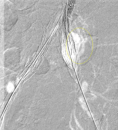
Every effort should be made to repair type I endoleak before completing the procedure (e.g., balloon molding of the proximal seal zone, placement of a proximal cuff, endostaples, liquid embolization).[206]van Schaik TG, Meekel JP, Hoksbergen AWJ, et al. Systematic review of embolization of type I endoleaks using liquid embolic agents. J Vasc Surg. 2021 Sep;74(3):1024-32.
https://www.doi.org/10.1016/j.jvs.2021.03.061
http://www.ncbi.nlm.nih.gov/pubmed/33940072?tool=bestpractice.com
Persistent type IA endoleak may necessitate conversion to open repair, provided the surgical risk is acceptable.[3]Wanhainen A, Van Herzeele I, Bastos Goncalves F, et al. Editor's choice -- European Society for Vascular Surgery (ESVS) 2024 clinical practice guidelines on the management of abdominal aorto-iliac artery aneurysms. Eur J Vasc Endovasc Surg. 2024 Feb;67(2):192-331.
https://www.ejves.com/article/S1078-5884(23)00889-4/fulltext
http://www.ncbi.nlm.nih.gov/pubmed/38307694?tool=bestpractice.com
[76]Chaikof EL, Dalman RL, Eskandari MK, et al. The Society for Vascular Surgery practice guidelines on the care of patients with an abdominal aortic aneurysm. J Vasc Surg. 2018 Jan;67(1):2-77.e2.
https://www.jvascsurg.org/article/S0741-5214(17)32369-8/fulltext
http://www.ncbi.nlm.nih.gov/pubmed/29268916?tool=bestpractice.com
[207]Perini P, Bianchini Massoni C, Mariani E, et al. Systematic review and meta-analysis of the outcome of different treatments for type 1a endoleak after EVAR. Ann Vasc Surg. 2019 Oct;60:435-46.e1.
http://www.ncbi.nlm.nih.gov/pubmed/31200054?tool=bestpractice.com
[Figure caption and citation for the preceding image starts]: Extension stent graft deployed for the same type I endoleak (encircled)University of Michigan/Dr G.R. Upchurch, Departments of Vascular Surgery and Radiology [Citation ends].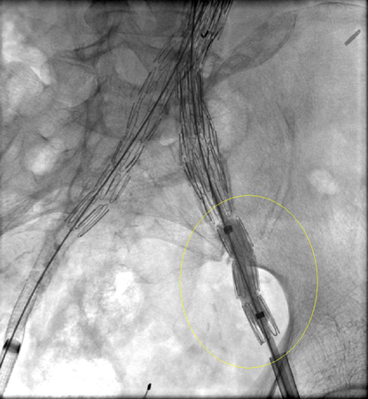 [Figure caption and citation for the preceding image starts]: Resolution of the type I endoleak resolved after extension deployedUniversity of Michigan/Dr G.R. Upchurch, Departments of Vascular Surgery and Radiology [Citation ends].
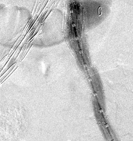
Type II:
Spontaneous resolution may occur, although persistence may result in sac growth.[208]Higashiura W, Greenberg RK, Katz E, et al. Predictive factors, morphologic effects, and proposed treatment paradigm for type II endoleaks after repair of infrarenal abdominal aortic aneurysms. J Vasc Interv Radiol. 2007 Aug;18(8):975-81.
http://www.ncbi.nlm.nih.gov/pubmed/17675614?tool=bestpractice.com
If a type II endoleak or other abnormality of concern is observed on contrast-enhanced CT imaging at 1 month after EVAR, postoperative imaging at 6 months is recommended.[76]Chaikof EL, Dalman RL, Eskandari MK, et al. The Society for Vascular Surgery practice guidelines on the care of patients with an abdominal aortic aneurysm. J Vasc Surg. 2018 Jan;67(1):2-77.e2.
https://www.jvascsurg.org/article/S0741-5214(17)32369-8/fulltext
http://www.ncbi.nlm.nih.gov/pubmed/29268916?tool=bestpractice.com
Around 50% of type II endoleaks are diagnosed before 30 day follow-up; 40% after 30 days, and 8% are diagnosed after 12 months of follow-up.[209]Charisis N, Bouris V, Conway AM, et al. A systematic review and pooled meta-analysis on the incidence and temporal occurrence of type II endoleak following an abdominal aortic aneurysm repair. Ann Vasc Surg. 2021 Aug;75:406-19.
http://www.ncbi.nlm.nih.gov/pubmed/33549794?tool=bestpractice.com
Treatment remains controversial and is advocated either if persistent at 6-12 months or when aneurysm sac size increases such that proximal and/or distal sealing zones may be compromised.[210]Mansueto G, Cenzi D, Scuro A, et al. Treatment of type II endoleak with a transcatheter transcaval approach: results at 1-year follow-up. J Vasc Surg. 2007 Jun;45(6):1120-7.
http://www.ncbi.nlm.nih.gov/pubmed/17543674?tool=bestpractice.com
[211]Baum RA, Stavropoulos SW, Fairman RM, et al. Endoleaks after endovascular repair of abdominal aortic aneurysms. J Vasc Interv Radiol. 2003 Sep;14(9 Pt 1):1111-7.
http://www.ncbi.nlm.nih.gov/pubmed/14514802?tool=bestpractice.com
[212]Van Marrewijk CJ, Fransen G, Laheij RJ, et al. Is a type II endoleak after EVAR a harbinger of risk? Causes and outcome of open conversion and aneurysm rupture during follow-up. Eur J Vasc Endovasc Surg. 2004 Feb;27(2):128-37.
http://www.ncbi.nlm.nih.gov/pubmed/14718893?tool=bestpractice.com
[213]Harris PL, Vallabhaneni SR, Desgranges P, et al. Incidence and risk factors of late rupture, conversion, and death after endovascular repair of infrarenal aortic aneurysms: the EUROSTAR experience. J Vasc Surg. 2000 Oct;32(4):739-49.
http://www.ncbi.nlm.nih.gov/pubmed/11013038?tool=bestpractice.com
[214]Ultee KHJ, Büttner S, Huurman R, et al. Editor's choice - systematic review and meta-analysis of the outcome of treatment for type II endoleak following endovascular aneurysm repair. Eur J Vasc Endovasc Surg. 2018 Dec;56(6):794-807.
https://www.doi.org/10.1016/j.ejvs.2018.06.009
http://www.ncbi.nlm.nih.gov/pubmed/30104089?tool=bestpractice.com
[215]Smith T, Quencer KB. Best practice guidelines: imaging surveillance after endovascular aneurysm repair. AJR Am J Roentgenol. 2020 May;214(5):1165-74.
https://www.doi.org/10.2214/AJR.19.22197
http://www.ncbi.nlm.nih.gov/pubmed/32130043?tool=bestpractice.com
Treatment of choice is transarterial coil embolization, although laparoscopic ligation of collateral branches, direct percutaneous translumbar puncture of the sac, translumbar embolization, and transcatheter transcaval embolization have been reported.[205]Veith FJ, Baum RA, Ohki T, et al. Nature and significance of endoleaks and endotension: summary of opinions expressed at an international conference. J Vasc Surg. 2002 May;35(5):1029-35.
http://www.ncbi.nlm.nih.gov/pubmed/12021724?tool=bestpractice.com
[210]Mansueto G, Cenzi D, Scuro A, et al. Treatment of type II endoleak with a transcatheter transcaval approach: results at 1-year follow-up. J Vasc Surg. 2007 Jun;45(6):1120-7.
http://www.ncbi.nlm.nih.gov/pubmed/17543674?tool=bestpractice.com
[211]Baum RA, Stavropoulos SW, Fairman RM, et al. Endoleaks after endovascular repair of abdominal aortic aneurysms. J Vasc Interv Radiol. 2003 Sep;14(9 Pt 1):1111-7.
http://www.ncbi.nlm.nih.gov/pubmed/14514802?tool=bestpractice.com
[212]Van Marrewijk CJ, Fransen G, Laheij RJ, et al. Is a type II endoleak after EVAR a harbinger of risk? Causes and outcome of open conversion and aneurysm rupture during follow-up. Eur J Vasc Endovasc Surg. 2004 Feb;27(2):128-37.
http://www.ncbi.nlm.nih.gov/pubmed/14718893?tool=bestpractice.com
[216]Steinmetz E, Rubin BG, Sanchez LA, et al. Type II endoleak after endovascular abdominal aortic aneurysm repair: a conservative approach with selective intervention is safe and cost-effective. J Vasc Surg. 2004 Feb;39(2):306-13.
http://www.ncbi.nlm.nih.gov/pubmed/14743129?tool=bestpractice.com
[217]Baum RA, Cope C, Fairman, et al. Translumbar embolization of type 2 endoleaks after endovascular repair of abdominal aortic aneurysms. J Vasc Interv Radiol. 2001 Jan;12(1):111-6.
http://www.ncbi.nlm.nih.gov/pubmed/11200344?tool=bestpractice.com
[218]Baum RA, Carpenter JP, Golden MA, et al. Treatment of type 2 endoleaks after endovascular repair of abdominal aortic aneurysms: comparison of transarterial and translumbar techniques. J Vasc Surg. 2002 Jan;35(1):23-9.
http://www.ncbi.nlm.nih.gov/pubmed/11802129?tool=bestpractice.com
[219]Schmid R, Gurke L, Aschwanden M, et al. CT-guided percutaneous embolization of a lumbar artery maintaining a type II endoleak. J Endovasc Ther. 2002 Apr;9(2):198-202.
http://www.ncbi.nlm.nih.gov/pubmed/12010100?tool=bestpractice.com
[220]Chin KM, Lee SQW, Lee HJ, et al. Preservation of stent graft after iatrogenic type III endoleak during open transperitoneal surgical intervention for complicated type II endoleak. Ann Vasc Surg. 2020 Jan;62:496.e1-496.e7.
http://www.ncbi.nlm.nih.gov/pubmed/31394250?tool=bestpractice.com
[221]Li Q, Hou P. Sac embolization and side branch embolization for preventing type II endoleaks after endovascular aneurysm repair: a meta-analysis. J Endovasc Ther. 2020 Feb;27(1):109-16.
https://www.doi.org/10.1177/1526602819878411
http://www.ncbi.nlm.nih.gov/pubmed/31566053?tool=bestpractice.com
[222]Akmal MM, Pabittei DR, Prapassaro T, et al. A systematic review of the current status of interventions for type II endoleak after EVAR for abdominal aortic aneurysms. Int J Surg. 2021 Nov;95:106138.
https://www.doi.org/10.1016/j.ijsu.2021.106138
http://www.ncbi.nlm.nih.gov/pubmed/34637951?tool=bestpractice.com
[223]Yu HYH, Lindström D, Wanhainen A, et al. Systematic review and meta-analysis of prophylactic aortic side branch embolization to prevent type II endoleaks. J Vasc Surg. 2020 Nov;72(5):1783-1792.e1.
https://www.doi.org/10.1016/j.jvs.2020.05.020
http://www.ncbi.nlm.nih.gov/pubmed/32442608?tool=bestpractice.com
[224]Zhang X, Ji L, Chen MY, et al. Transarterial embolization versus translumber embolization for type Ⅱ endoleak after endovascular abdominal aortic aneurysm repair: a meta-analysis. Chin Med Sci J. 2020 Jun 30;35(2):135-41.
https://www.doi.org/10.24920/003618
http://www.ncbi.nlm.nih.gov/pubmed/32684233?tool=bestpractice.com
Type III:
Graft defect with leak through fabric tears, graft disconnection, or disintegration of the fabric.[204]Schurink GW, Aarts NJ, vanBockel JH. Endoleak after stent-graft treatment of abdominal aortic aneurysm: a meta-analysis of clinical studies. Br J Surg. 1999 May;86(5):581-7.
http://www.ncbi.nlm.nih.gov/pubmed/10361173?tool=bestpractice.com
[205]Veith FJ, Baum RA, Ohki T, et al. Nature and significance of endoleaks and endotension: summary of opinions expressed at an international conference. J Vasc Surg. 2002 May;35(5):1029-35.
http://www.ncbi.nlm.nih.gov/pubmed/12021724?tool=bestpractice.com
[225]Kwon J, Dimuzio P, Salvatore D, et al. Incidence of stent graft failure from type IIIB endoleak in contemporary endovascular abdominal aortic aneurysm repair. J Vasc Surg. 2020 Feb;71(2):645-53.
https://www.doi.org/10.1016/j.jvs.2019.06.183
http://www.ncbi.nlm.nih.gov/pubmed/31466740?tool=bestpractice.com
Repair is indicated upon discovery (endovascular stent graft extension).[3]Wanhainen A, Van Herzeele I, Bastos Goncalves F, et al. Editor's choice -- European Society for Vascular Surgery (ESVS) 2024 clinical practice guidelines on the management of abdominal aorto-iliac artery aneurysms. Eur J Vasc Endovasc Surg. 2024 Feb;67(2):192-331.
https://www.ejves.com/article/S1078-5884(23)00889-4/fulltext
http://www.ncbi.nlm.nih.gov/pubmed/38307694?tool=bestpractice.com
[76]Chaikof EL, Dalman RL, Eskandari MK, et al. The Society for Vascular Surgery practice guidelines on the care of patients with an abdominal aortic aneurysm. J Vasc Surg. 2018 Jan;67(1):2-77.e2.
https://www.jvascsurg.org/article/S0741-5214(17)32369-8/fulltext
http://www.ncbi.nlm.nih.gov/pubmed/29268916?tool=bestpractice.com
[211]Baum RA, Stavropoulos SW, Fairman RM, et al. Endoleaks after endovascular repair of abdominal aortic aneurysms. J Vasc Interv Radiol. 2003 Sep;14(9 Pt 1):1111-7.
http://www.ncbi.nlm.nih.gov/pubmed/14514802?tool=bestpractice.com
[215]Smith T, Quencer KB. Best practice guidelines: imaging surveillance after endovascular aneurysm repair. AJR Am J Roentgenol. 2020 May;214(5):1165-74.
https://www.doi.org/10.2214/AJR.19.22197
http://www.ncbi.nlm.nih.gov/pubmed/32130043?tool=bestpractice.com
[226]Lowe C, Hansrani V, Madan M, et al. Type IIIb endoleak after elective endovascular aneurysm repair: a systematic review. J Cardiovasc Surg (Torino). 2020 Jun;61(3):308-16.
http://www.ncbi.nlm.nih.gov/pubmed/29616524?tool=bestpractice.com
Type IV:
Leak from graft wall porosity.[204]Schurink GW, Aarts NJ, vanBockel JH. Endoleak after stent-graft treatment of abdominal aortic aneurysm: a meta-analysis of clinical studies. Br J Surg. 1999 May;86(5):581-7.
http://www.ncbi.nlm.nih.gov/pubmed/10361173?tool=bestpractice.com
[205]Veith FJ, Baum RA, Ohki T, et al. Nature and significance of endoleaks and endotension: summary of opinions expressed at an international conference. J Vasc Surg. 2002 May;35(5):1029-35.
http://www.ncbi.nlm.nih.gov/pubmed/12021724?tool=bestpractice.com
These leaks are uncommon with newer stent grafts and are self-limited.[76]Chaikof EL, Dalman RL, Eskandari MK, et al. The Society for Vascular Surgery practice guidelines on the care of patients with an abdominal aortic aneurysm. J Vasc Surg. 2018 Jan;67(1):2-77.e2.
https://www.jvascsurg.org/article/S0741-5214(17)32369-8/fulltext
http://www.ncbi.nlm.nih.gov/pubmed/29268916?tool=bestpractice.com
[211]Baum RA, Stavropoulos SW, Fairman RM, et al. Endoleaks after endovascular repair of abdominal aortic aneurysms. J Vasc Interv Radiol. 2003 Sep;14(9 Pt 1):1111-7.
http://www.ncbi.nlm.nih.gov/pubmed/14514802?tool=bestpractice.com
Type V (endotension):
Endotension is increased intrasac pressure after EVAR without visualized endoleak on delayed contrast CT scans.
Endotension is less common with the newer-generation grafts.[76]Chaikof EL, Dalman RL, Eskandari MK, et al. The Society for Vascular Surgery practice guidelines on the care of patients with an abdominal aortic aneurysm. J Vasc Surg. 2018 Jan;67(1):2-77.e2.
https://www.jvascsurg.org/article/S0741-5214(17)32369-8/fulltext
http://www.ncbi.nlm.nih.gov/pubmed/29268916?tool=bestpractice.com
There is no standardized method to measure endotension or consensus on indicated therapy in the absence of aneurysm enlargement; however, treatment of endotension to prevent aneurysm rupture is suggested in selected patients with continued aneurysm expansion.[76]Chaikof EL, Dalman RL, Eskandari MK, et al. The Society for Vascular Surgery practice guidelines on the care of patients with an abdominal aortic aneurysm. J Vasc Surg. 2018 Jan;67(1):2-77.e2.
https://www.jvascsurg.org/article/S0741-5214(17)32369-8/fulltext
http://www.ncbi.nlm.nih.gov/pubmed/29268916?tool=bestpractice.com
[205]Veith FJ, Baum RA, Ohki T, et al. Nature and significance of endoleaks and endotension: summary of opinions expressed at an international conference. J Vasc Surg. 2002 May;35(5):1029-35.
http://www.ncbi.nlm.nih.gov/pubmed/12021724?tool=bestpractice.com
[215]Smith T, Quencer KB. Best practice guidelines: imaging surveillance after endovascular aneurysm repair. AJR Am J Roentgenol. 2020 May;214(5):1165-74.
https://www.doi.org/10.2214/AJR.19.22197
http://www.ncbi.nlm.nih.gov/pubmed/32130043?tool=bestpractice.com