Approach
Hyponatremia is essentially a laboratory diagnosis, defined as a serum sodium concentration of <135 mEq/L.[2][3] History and physical exam establish volume status and are used to determine if the patient is hypovolemic, hypervolemic, or euvolemic. A thorough review of any underlying medical conditions and medications should be undertaken. The cause of the hyponatremia is often apparent from the history and exam; however, some causes can only be identified with appropriate investigations. Patients with asymptomatic, mild hyponatremia (130-135 mEq/L) may be managed initially in a community setting.[41] Initial assessment should also include glycemic status, to establish whether there is a hyperglycemic-induced hyponatremia, and the exclusion of pseudohyponatremia caused by excessive plasma lipids or proteins. Patients with onset of hyponatremia in <48 hours, or those with symptoms, require urgent assessment.
History
Hyponatremia can be present on admission to hospital, or it can develop (or worsen) during the hospital stay as a result of several factors including organ failure, medications, or the postoperative state.[2][11][24]
Patients should be asked about fluid intake.
Hypovolemic hyponatremia:
Recent excessive fluid losses should be noted (e.g., severe diarrhea or vomiting), which may point to hypovolemic hyponatremia. Third spacing of fluids (e.g., in pancreatitis or severe hypoalbuminemia) may also lead to hypovolemic hyponatremia. A history of diabetes mellitus should be sought.
Hypervolemic hyponatremia:
Typically associated with congestive heart failure, cirrhosis, nephrotic syndrome, or acute kidney injury/chronic kidney disease.
Euvolemic hyponatremia:
May result from excessive fluid intake, as might occur during high-intensity physical exercise (e.g., marathon running, military training, wilderness exploration).[16][40] Such intake may be associated with acute, symptomatic hyponatremia.[40]
Potomania is caused by high fluid intake in the setting of very low solute and electrolyte intake. Maximal urinary dilution may be impaired by very low protein intake, and it also reduces with advancing age. It can occur in association with inadequate diets (i.e., very low-calorie diets with high fluid intake, such as the "tea and toast" diet), crash diets and alcohol use disorder with a high intake of beer. If a history of alcohol use disorder is present or suspected, the CAGE questionnaire can be used. MDCalc: CAGE questions for alcohol use Opens in new window A score >2 is considered indicative of probable excessive alcohol intake.
Although less common, euvolemic hyponatremia related to excessive fluids can occur in the setting of surgery or medical testing such as cardiac catheterization or colonoscopy.
Pseudohyponatremia:
Suspected in settings such as history of multiple myeloma (due to high serum protein levels) or severe hyperlipidemia.
A drug history should be taken, since a number of medications are associated with hyponatremia including:
Vasopressin analogs: desmopressin, oxytocin
Medications that stimulate vasopressin release or potentiate the effects of vasopressin: selective serotonin-reuptake inhibitors and most other antidepressants, morphine and other opioids[18][19]
Medications that impair urinary dilution: thiazide diuretics[12][15][20]
Medications that cause hyponatremia by an uncertain mechanism of action: carbamazepine or its analogs, vincristine, nicotine, antipsychotics, chlorpropamide, cyclophosphamide, nonsteroidal anti-inflammatory drugs[17]
Illicit drugs: methylenedioxy-methamphetamine (MDMA or ecstasy) causes vasopressin release, and has been associated with acute, life-threatening hyponatremia
Syndrome of inappropriate antidiuretic hormone (SIADH) is a common cause of euvolemic hyponatremia, and is a diagnosis of exclusion. Hypothyroidism and adrenal insufficiency must be excluded for a diagnosis of SIADH to be made.[4] SIADH is associated with many lung diseases, particularly those resulting from infection such as pneumonia; central nervous system diseases, such as subarachnoid hemorrhage, meningitis, or encephalitis; or malignancies, most commonly small cell lung cancer, or gastrointestinal tract cancers.[4] It can also be due to other nonspecific causes (e.g., drugs, pain, nausea, stress, general anesthesia), and an idiopathic form of SIADH that is associated with aging may occur.[23][31] At times, SIADH may be the presenting finding of a malignancy, or it may precede the malignancy diagnosis. If there is no identifiable cause, the patient can be diagnosed as having idiopathic SIADH.[4]
If hyponatremia develops acutely (i.e., <48 hours), the brain does not have time to adapt and cerebral edema occurs, leading to symptoms including nausea, vomiting, altered mental status, and eventually seizures and/or brain herniation and death. This is a medical emergency requiring hypertonic 3% saline for management.[5] With more chronic hyponatremia, brain adaptation occurs with the loss of intracellular osmolytes, returning brain volume to normal after approximately 48 hours. These patients are generally asymptomatic or present with only mild cognitive symptoms (e.g., confusion, balance difficulties).
If a patient presents with acute hyponatremia and a history of altered mental status (e.g., schizophrenia or psychotic depression) with seizures, a diagnosis of primary polydipsia should be considered.
Physical exam
The patient should be examined to determine volume status, looking for signs of dehydration or volume overload.[3]
Signs of volume depletion include:
Low urine output
Weight loss
Orthostatic hypotension
Decreased jugular venous pressure
Poor skin turgor
Dry mucus membranes
Absence of axillary sweat
Absence of edema
Signs of volume overload include:
Edema and/or ascites
Rales or crackles on lung auscultation
Significant weight gain
Raised jugular venous pressure
Polyuria is most common in primary polydipsia.
At times the physical exam and vital signs may be normal in patients with hypovolemic hyponatremia; therefore, the cause may not initially be clear.[3] Further investigations (e.g., urine electrolytes, fractional excretion of sodium) aid diagnosis. Patients with hypervolemic hyponatremia typically have signs and symptoms that point to a diagnosis of heart failure, cirrhosis, or nephrotic syndrome, although kidney disease should also be considered.
Exclusion of hypertonic hyponatremia or pseudohyponatremia
Serum glucose (random or fasting) should be checked to exclude hyperglycemia-associated hyponatremia. If the patient is hyperglycemic, a sodium correction formula should be used if the glucose level is >100 mg/dL.
The most accurate correction formula is:
Corrected serum sodium (mEq/L) = measured serum sodium (mEq/L) + 2.4 × {[serum glucose (mg/dL) - 100]/100} [ Sodium Correction in Hyperglycemia (Hillier 1999) Opens in new window ]
This formula should be used to determine if true hyponatremia is present.[42] It is important to remember that even in the setting of hyperglycemia, true hyponatremia may occur. The corrected sodium level may be low (hypotonic hyponatremia), normal (isotonic hyponatremia), or high (hypertonic hyponatremia). For example, in diabetic ketoacidosis, urine losses may be isotonic and water intake may be high leading to an underlying hypotonic hyponatremia that is unmasked once glucose levels are lowered with insulin. Similarly, a patient with uncontrolled diabetes mellitus and congestive heart failure may have a low serum sodium concentration once the hyperglycemia is corrected.
To exclude the possibility of pseudohyponatremia, blood lipids and proteins should be measured before proceeding with further investigations. Serum osmolality will be normal in patients with pseudohyponatremia (i.e., total body sodium and water are unchanged, with no shift of fluid between intracellular and extracellular compartments). The use of ion-specific electrodes to measure sodium directly may help reduce the incidence of pseudohyponatremia.
Investigations
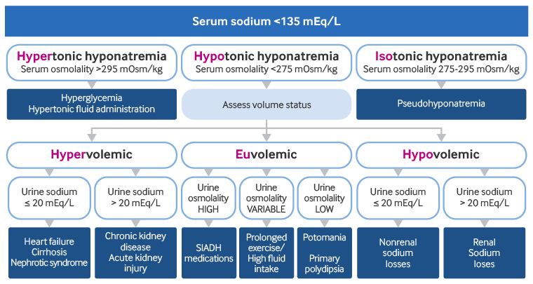
[Figure caption and citation for the preceding image starts]: Algorithm for the diagnosis of hypotonic hyponatremia. SIADH, syndrome of inappropriate antidiuretic hormoneCreated by BMJ Knowledge Centre [Citation ends].
A full serum electrolyte panel with glucose, blood urea nitrogen, and creatinine should be ordered in all patients. A serum sodium concentration <135 mEq/L (corrected for hyperglycemia) confirms the presence of hyponatremia.
The following tests should also be ordered in all patients (preferably before the administration of intravenous fluids) to help establish the underlying etiology:
Serum osmolality
Urine sodium and creatinine concentrations
Urine osmolality
Serum osmolality
Serum osmolality can differentiate between hypotonic, hypertonic, and isotonic hyponatremia:[2][3] [ Osmolality Estimator (serum) Opens in new window ]
Serum osmolality <275 mOsm/kg: indicates hypotonic hyponatremia
Serum osmolality >295 mOsm/kg: indicates hypertonic hyponatremia
Serum osmolality normal: indicates isotonic hyponatremia (pseudohyponatremia)
Hypertonic hyponatremia is due to either hyperglycemia or the administration of hypertonic fluids (e.g., mannitol, sorbitol). Isotonic hyponatremia indicates pseudohyponatremia. Hypotonic hyponatremia has a wider range of causes, and can be further classified as:
Hypovolemic: clinical features of volume depletion present
Hypervolemic: clinical features of fluid overload present
Euvolemic: absence of signs of volume depletion or overload
Urine sodium concentration
It may be difficult to distinguish between hypovolemia and euvolemia on physical exam. A spot urine test allows urinary sodium concentration to be measured quickly and conveniently and may help confirm the presence of hypovolemia or euvolemia.
The urine sodium concentration, in combination with the volume status of the patient from exam, can provide further clues to the classification and etiology.
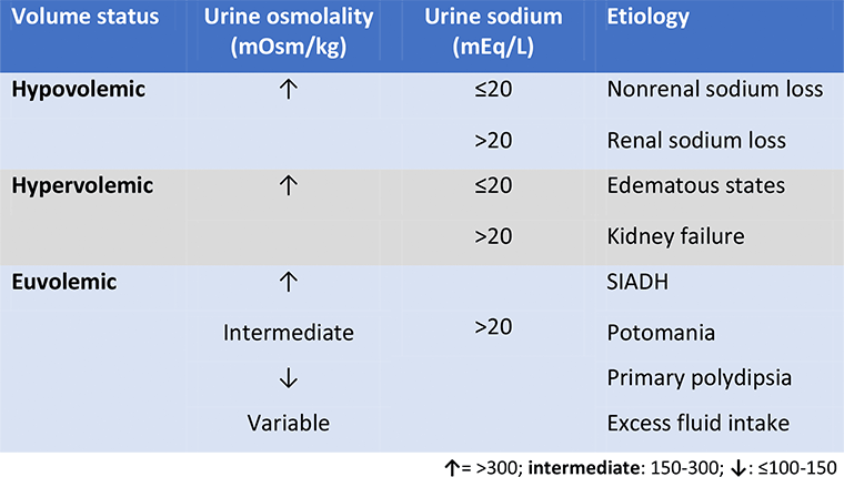
[Figure caption and citation for the preceding image starts]: Etiologies of hypotonic hyponatremia (serum osmolality <275 mOsm/kg). SIADH, syndrome of inappropriate antidiuretic hormoneProduced by the BMJ Knowledge Centre [Citation ends].
Hypovolemic hyponatremia:
Urine sodium concentration >20 mEq/L: indicates renal sodium losses (e.g., diuretics)
Urine sodium concentration ≤20 mEq/L: indicates hypovolemia with nonrenal sodium losses (e.g., gastrointestinal losses)
Hypervolemic hyponatremia:
Urine sodium concentration >20 mEq/L: indicates acute kidney injury/chronic kidney disease or diuretic use
Urine sodium concentration ≤20 mEq/L: indicates congestive heart failure, cirrhosis, or nephrotic syndrome
Euvolemic hyponatremia:
Urine sodium concentration is >20 mEq/L in most patients with euvolemic hyponatremia; however, patients with a concomitant low sodium intake may have a low urinary sodium.
Although a spot urine sodium test can be helpful if the result is very low, the fractional excretion of sodium provides a more accurate assessment of volume status as it corrects for the effect of variations in urine volume on the urine sodium. [ Fractional Excretion of Sodium Opens in new window ] The fractional excretion of sodium is calculated using the following formula:
[(urinary sodium concentration × plasma creatinine concentration)/(plasma sodium concentration × urinary creatinine concentration)] × 100% [ Fractional Excretion of Sodium Opens in new window ]
A value of <1% usually indicates pre-renal causes of hyponatremia.
Urine osmolality
Urine osmolality can be used to further evaluate the cause of hyponatremia in patients, especially those with euvolemic hyponatremia. Urine osmolality is high (i.e., ≥300 mOsm/kg) in patients with hypovolemic or hypervolemic hyponatremia, but can be variable in patients with euvolemic hyponatremia:[5]
High (≥300 mOsm/kg): indicates SIADH due to the inappropriate dilution of plasma as a result of pathologic vasopressin release, or may be due to drug-related effects.[2]
Intermediate (150-300 mOsm/kg): indicates potomania or a partial effect of medications or mild SIADH in conjunction with high fluid intake.
Low (≤100-150 mOsm/kg): indicates primary polydipsia.
Urine osmolality can be variable in hyponatremia due to prolonged physical exercise with high fluid intake. This is because it reflects vasopressin release with high urine osmolality initially, followed by low urine osmolality as self-correction and aquaresis (loss of water) occur.
Electrolyte-free water excretion
Urine electrolytes and urine flow rate are used to calculate the electrolyte-free water excretion (also known as electrolyte-free water clearance). This value helps to determine the treatment plan by determining ongoing water gains or losses.
It should be calculated in patients with hyponatremia using the following formula:
Electrolyte-free water excretion = V × [1 - (UNa + UK)/(PNa)]
Where V is the urine flow rate, UNa is the urine concentration of sodium (mEq/L), UK is the urine concentration of potassium (mEq/L), and PNa is the plasma concentration of sodium (mEq/L).
The resulting value indicates how much electrolyte-free water is being lost through the urine at any given time (e.g., per hour, per day). This formula can be used to help determine if water is being retained (a negative value) as in SIADH, or excreted (positive value) as in polydipsia or potomania.[43][44]
Other investigations
Thyroid function tests should be ordered to exclude hypothyroidism, and serum cortisol level adrenocorticotropic hormone testing to exclude adrenal insufficiency in patients with euvolemic hyponatremia. Hypothyroidism and adrenal insufficiency must be excluded for a diagnosis of SIADH to be made.[4]
A computed tomography scan of the brain, chest, and/or abdomen/pelvis should be ordered to identify potential causes of SIADH and should be guided by history and physical exam.[4] At times, SIADH may be the presenting finding of a malignancy, or it may precede the malignancy diagnosis.
Other tests targeted at diagnosing the underlying cause may be required.
Use of this content is subject to our disclaimer