Images and videos
Images


Evaluation of ptosis
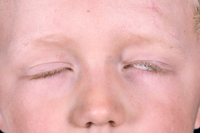
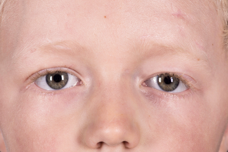
Ptosis in a 6-year-old boy. Ptosis is normally due to weakness of the levator muscle of the upper eyelid, here of the left eye (at right). This patient has had this condition since birth, and has had three operations aimed at correcting the condition
Mid Essex Hospital Services NHS Trust/Science Photo Library; used with permission
See this image in context in the following section/s:
Evaluation of ptosis
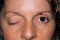
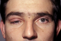
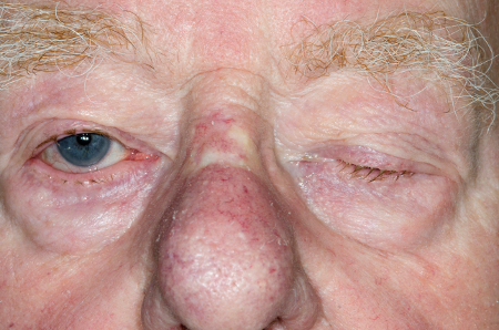
Oculomotor nerve palsy. Face of a 36-year-old woman with third nerve palsy after surgery to treat a subarachnoid haemorrhage. A berry aneurysm, a common localised dilation of an intercranial artery, caused the subarachnoid hemorrhage. Third nerve palsy is a dysfunction of the third cranial nerve, the oculomotor nerve, which controls the movement of the eyes. It leads to an inability to move the eye, double vision, a fixed and non-reactive pupil and eyelid drooping (ptosis, seen here, right eye)
Dr P Marazzi/Science Photo Library; used with permission
See this image in context in the following section/s:

Evaluation of ptosis
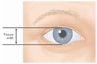
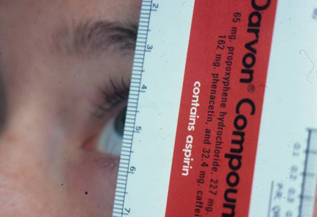
Measurement of vertical interpalpebral fissure
From the collection of Dr Allen Putterman
See this image in context in the following section/s:
Evaluation of ptosis
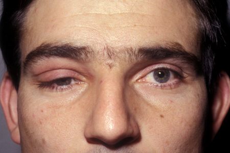
Male patient suffering from post-traumatic acquired ptosis, likely caused by an injury to the eyelid
Barraquer, Barcelona - ISM/Science Photo Library; used with permission
See this image in context in the following section/s:
Evaluation of ptosis
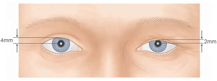
Measurement of margin-reflex distance
From the collection of Dr Allen Putterman
See this image in context in the following section/s:
Evaluation of ptosis
Ptosis in an 89 year old male patient following a botox injection to correct double vision (diplopia)
Dr P Marazzi/Science Photo Library; used with permission
See this image in context in the following section/s:

Evaluation of ptosis
Oculomotor nerve palsy. Face of a 36-year-old woman with third nerve palsy after surgery to treat a subarachnoid haemorrhage. A berry aneurysm, a common localised dilation of an intercranial artery, caused the subarachnoid hemorrhage. Third nerve palsy is a dysfunction of the third cranial nerve, the oculomotor nerve, which controls the movement of the eyes. It leads to an inability to move the eye, double vision, a fixed and non-reactive pupil and eyelid drooping (ptosis, seen here, right eye)
Dr P Marazzi/Science Photo Library; used with permission
See this image in context in the following section/s:

Evaluation of ptosis
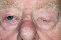
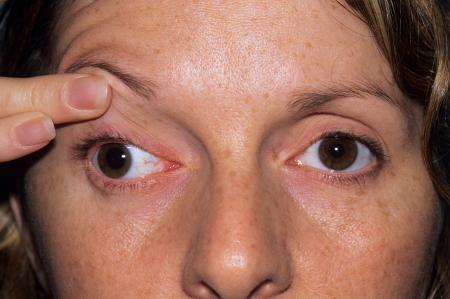
Drooping eyelid (ptosis) in 69 year old female patient due to myasthenia gravis (MG). MG is a rare autoimmune neuromuscular disorder that weakens and fatigues the body's voluntary muscles, which include the muscles that control movement of the eyes and eyelids
Dr P Marazzi/Science Photo Library; used with permission
See this image in context in the following section/s:

Evaluation of ptosis
Position of upper eyelid in downgaze
From the collection of Dr Allen Putterman
See this image in context in the following section/s:
Evaluation of ptosis
Sagittal view of eyelid anatomy
From the collection of Dr Allen Putterman
See this image in context in the following section/s:
Evaluation of ptosis
Position of upper eyelid in upgaze
From the collection of Dr Allen Putterman
See this image in context in the following section/s:
Use of this content is subject to our disclaimer