Approach
Risk factors related to the development of abdominal wall defects are generally thought to be unmodifiable. However, maternal health factors associated with the development of abdominal wall defects include smoking and the use of certain recreational drugs.[3][4][9][10] There are strong associations related to the discovery of these abdominal wall defects, which include maternal age <20 years for the development of gastroschisis and maternal age >40 years for omphalocele. Additionally, gastroschisis is more likely to be seen in male infants than in female infants. Weak factors associated with the development of abdominal wall defects include infections during pregnancy.
Abdominal wall defects of the fetus are routinely detected on prenatal ultrasound in the second trimester, when an abdominal mass may be visualized outside of the abdominal wall. Elevated alpha-fetoprotein levels, measured as part of the routine maternal triple screen at 16 to 18 weeks' gestation, suggest the presence of an abdominal wall defect and warrant further investigation with prenatal ultrasonography.[21] Definitive diagnosis is achieved at birth through a careful physical exam of the infant revealing abdominal contents external to the abdominal wall.
Detection of abdominal wall defects in the prenatal period allows parental counseling and the development of a postdelivery treatment plan, as well as arrangements for the delivery to take place at a tertiary care center equipped to handle the resuscitative and surgical needs of the newborn infant.[9][17][20][22][23]
Prenatal assessment
Elevated maternal serum alpha-fetoprotein levels reflect protein loss from the intestine floating in the amniotic fluid, and are a reliable but nonspecific marker for the presence of gastroschisis. Abnormal maternal serum screening results warrant additional imaging to confirm the diagnosis of an abdominal wall defect, with high-definition ultrasound in addition to routine abdominal ultrasonography to visualize the developing fetus.
If evidence of omphalocele is confirmed on ultrasonography, invasive procedures such as amniocentesis performed at 15 to 20 weeks' gestation or chorionic villus sampling performed at 10 to 12 weeks' gestation may be undertaken to evaluate the possibility of associated chromosomal abnormalities, such as trisomy 13, trisomy 18, trisomy 21, Turner syndrome, Klinefelter syndrome, or triploidy.[9][17][20] Chromosomal abnormalities are more common in omphalocele than gastroschisis.
In many pregnancies, despite normal serum screening results, an abdominal wall defect may be detected on a routine prenatal ultrasound. During second-trimester ultrasonography, these defects are characterized by abdominal wall masses and echogenic bowel outside of the abdominal wall.
As associated genetic anomalies can occur in as many as 30% to 40% of infants with prenatal evidence of omphalocele, prenatal genetic testing for chromosomal abnormalities, and an echocardiogram for detection of cardiac anomalies, are routinely performed.[24][25] Infants with prenatal evidence of gastroschisis do not routinely receive prenatal genetic testing.
In gastroschisis, dilated, fluid-filled loops of bowel floating freely in the amniotic fluid, and in some cases intestinal atresia, may be detected on ultrasound. If omphalocele is suspected, sternal and cardiac defects should be evaluated on ultrasound to determine the potential presence of pentalogy of Cantrell and Beckwith-Wiedemann syndrome, the latter being confirmed by the presence of macroglossia and visceromegaly.[17][22]
When an abdominal wall defect is visualized on ultrasound, in addition to the diagnosis of gastroschisis or omphalocele, it is important to consider the possibility of cloacal exstrophy, which is characterized by a portion of the large intestine lying outside of the body and splitting the bladder into two halves. In boys, the penis is usually flat and short, with the exposed inner surface of the urethra on top. The penis is sometimes split into a right and left half. In girls, the clitoris is split and there may be one or two vaginal openings. Cloacal exstrophy is a very rare birth defect, affecting 1 in every 200,000 to 400,000 births.[26]
The optimal mode of delivery of infants with a prenatally diagnosed abdominal wall defect is intensely controversial.[17] Anecdotal evidence suggests that vaginal delivery may be contraindicated for infants with abdominal wall defects, particularly gastroschisis. However, other reports have found that avoiding vaginal delivery for these infants confers no benefit.[23][27][28][29][30]
Postnatal exam
A careful physical exam at birth revealing abdominal contents external to the abdominal wall is diagnostic of omphalocele and gastroschisis.
Omphaloceles are abdominal wall defects ranging from 4 to 12 cm in size and can be located centrally, or in the epigastric or hypogastric regions. In omphalocele, as the abdominal contents have a protective membranous covering in utero, the intestines are usually healthy at birth. This condition may be associated with Beckwith-Wiedemann syndrome and, rarely, with pentalogy of Cantrell. The likelihood of additional developmental abnormalities, most often affecting cardiac anatomy, is high, leading to the descriptive phrase "bad baby, good bowel." Prenatal ultrasonography and postnatal physical exam will detect most of the developmental abnormalities associated with omphalocele.
In gastroschisis the lack of a protective membranous covering causes the abdominal contents to be free-floating in utero. This leads to a chemical reaction that creates a thick inflammatory film or peel overlying the intestine. Gastroschisis is commonly associated with intestinal atresia, which occurs in 10% to 15% of infants, and is related to ischemia of the exposed gut caused by constriction of its mesenteric blood supply at the level of the abdominal wall defect.[31] There are few additional structural abnormalities, although the intestine may require a lengthy period to recover, leading to the descriptive phrase "good baby, bad bowel." [Figure caption and citation for the preceding image starts]: Note the membrane covering the abdominal contents in this omphaloceleFrom collection of J.J. Tepas III, MD, FACS, FAAP [Citation ends].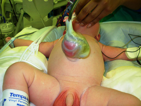 [Figure caption and citation for the preceding image starts]: Extruded gut in abdominal wall defectFrom collection of J.J. Tepas III, MD, FACS, FAAP [Citation ends].
Investigations
During the neonatal period, infants with omphalocele and gastroschisis do not routinely receive any further investigations unless there are signs of dysmorphia. However, genetic counseling is routinely offered because many families request assistance in determining future child-bearing plans.
Use of this content is subject to our disclaimer