Images and videos
Images

Eustachian tube dysfunction
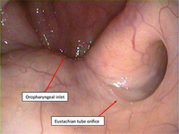
A normal left Eustachian tube as seen endoscopically from the posterior nasal cavity.
From the collection of Edward D. McCoul; used with permission
See this image in context in the following section/s:
Eustachian tube dysfunction
Type C tympanogram, demonstrating a malfunctioning Eustachian tube
From the collection of Erica R. Thaler; used with permission
See this image in context in the following section/s:

Eustachian tube dysfunction
Normal (type A) tympanogram
From the collection of Erica R. Thaler; used with permission
See this image in context in the following section/s:

Eustachian tube dysfunction
Type B tympanogram; flat compliance curve demonstrates no movement of the tympanic membrane
From the collection of Erica R. Thaler; used with permission
See this image in context in the following section/s:
Eustachian tube dysfunction
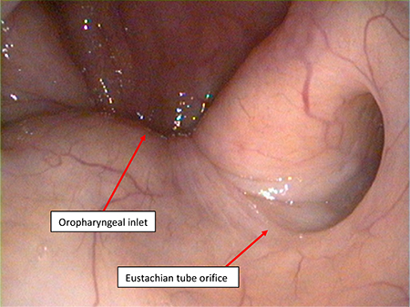
A normal left Eustachian tube as seen endoscopically from the posterior nasal cavity.
From the collection of Edward D. McCoul; used with permission
See this image in context in the following section/s:
Use of this content is subject to our disclaimer