Images and videos
Images


Cerebral arteriovenous malformation
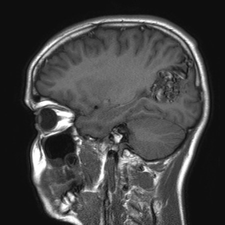
Unruptured left parieto-occipital arteriovenous malformation (sagittal T1-weighted magnetic resonance imaging scan)
From the collection of Mr R. J. Edwards; used with permission
See this image in context in the following section/s:

Cerebral arteriovenous malformation
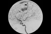
Cerebral angiogram (left carotid artery injection, lateral view) showing posterior frontal arteriovenous malformation fed by pericallosal artery (thin arrow) with arterialized draining vein (thick arrow) draining to superior sagittal sinus
From the collection of Mr R. J. Edwards; used with permission
See this image in context in the following section/s:
Cerebral arteriovenous malformation
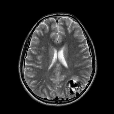
Unruptured left parieto-occipital arteriovenous malformation (axial T2-weighted magnetic resonance imaging scan)
From the collection of Mr R. J. Edwards; used with permission
See this image in context in the following section/s:
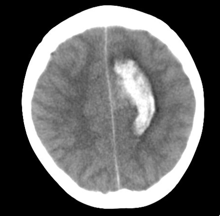
Cerebral arteriovenous malformation
Left posterior frontal intracerebral hematoma secondary to ruptured arteriovenous malformation (axial unenhanced computed tomography scan)
From the collection of Mr R. J. Edwards; used with permission
See this image in context in the following section/s:
Cerebral arteriovenous malformation
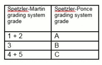
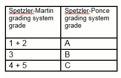
Consolidation of the Spetzler-Martin grading system into the Spetzler-Ponce grading system
Created by the BMJ Knowledge Centre
See this image in context in the following section/s:
Use of this content is subject to our disclaimer