Images and videos
Images


Oropharyngeal cancer
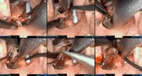
Robotic wide-field tonsillectomy using the Da Vinci SP robot
From the collection of Dr Linda X. Yin; used with permission
See this image in context in the following section/s:

Oropharyngeal cancer
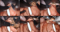
Robotic base of tongue resection using the Da Vinci SP robot
From the collection of Dr Linda X. Yin; used with permission
See this image in context in the following section/s:

Oropharyngeal cancer
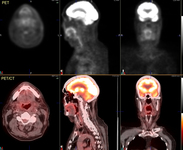
60-year-old man with squamous cell carcinoma of the right tongue base. PET/CT images show mild increased metabolism in a mid right neck lymph node, of concern for metastatic involvement (arrows)
From the collection of Dr Fabio Almeida; used with permission
See this image in context in the following section/s:

Oropharyngeal cancer
Large base of tongue tumor not amenable to transoral robotic surgery, required open transhyoid approach to the base of tongue
From the collection of Dr Linda X. Yin; used with permission
See this image in context in the following section/s:
Oropharyngeal cancer
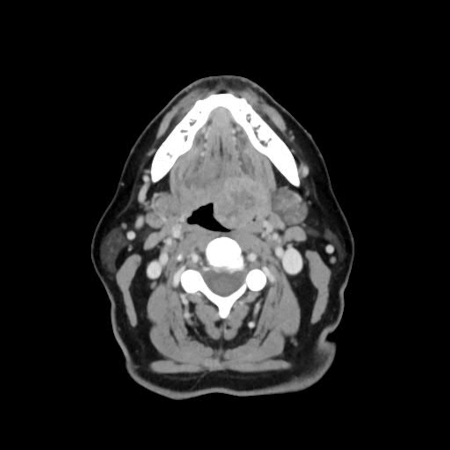
Large base of tongue tumor seen on axial CT scan
From the collection of Dr Linda X. Yin; used with permission
See this image in context in the following section/s:

Oropharyngeal cancer
74-year-old man with squamous cell carcinoma of the left tongue base extending into the hypopharynx. Fluorodeoxyglucose PET/CT images demonstrate focal increased metabolic activity in the left hypopharynx/tongue base (arrows)
From the collection of Dr Fabio Almeida; used with permission
See this image in context in the following section/s:
Oropharyngeal cancer
74-year-old man with squamous cell carcinoma of the left tongue base extending into the hypopharynx. Images after chemoradiation therapy, showing complete resolution of metabolic foci. Mild diffuse increased metabolism in the oropharyngeal region consistent with mild post-therapy inflammation
From the collection of Dr Fabio Almeida; used with permission
See this image in context in the following section/s:
Oropharyngeal cancer
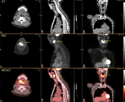
60-year-old man with squamous cell carcinoma of the right tongue base. Axial images further caudally show extent of tumor involvement in the hypopharynx including invasion through the hyoid bone
From the collection of Dr Fabio Almeida; used with permission
See this image in context in the following section/s:

Oropharyngeal cancer
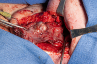
Transhyoid open approach to a large base of tongue tumor
From the collection of Dr Linda X. Yin; used with permission
See this image in context in the following section/s:

Oropharyngeal cancer
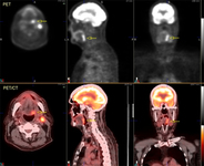
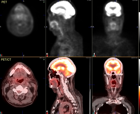
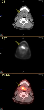
60-year-old man with squamous cell carcinoma of the right tongue base. Fluorodeoxyglucose PET/CT images demonstrate focal increased metabolic activity in the right tongue base, which extends inferior to the hypopharynx (arrows) and across the midline. On the CT images (top row), soft-tissue irregularity can be seen, but the margins of the tumor are difficult to define
From the collection of Dr Fabio Almeida; used with permission
See this image in context in the following section/s:
Use of this content is subject to our disclaimer