Tests
1st tests to order
chest x-ray (posteroanterior and lateral)
Test
Initial screening test in an individual with exposure to silica or coal. A blood beryllium lymphocyte proliferation test is the initial screening test in an individual exposed to beryllium.
Less sensitive than high-resolution CT (HRCT) scan, and more specific than pulmonary function testing (spirometry, lung volumes, and diffusing capacity).
The presence of noncalcified, multiple (in the hundreds), rounded opacities in the upper zones is highly suggestive of silicosis or coal workers' pneumoconiosis. Progressively involves all lung zones, and may conglomerate to form large opacities. With advanced conglomeration, there may be distortion of lung and heart shape, and a thin layer of calcification around hilar lymph nodes ("egg shell calcification").
Although the usual radiographic changes with silicosis and coal workers' pneumoconiosis are rounded opacities, linear changes have been described.[39]
Similarly, although the usual radiographic changes with chronic beryllium disease are linear, rounded opacities have been described.[40][Figure caption and citation for the preceding image starts]: CXR showing changes consistent with simple silicosis or coal workers' pneumoconiosisFrom the personal collection of Kenneth D. Rosenman, Michigan State University [Citation ends]. [Figure caption and citation for the preceding image starts]: CXR of chronic beryllium diseaseFrom the personal collection of Kenneth D. Rosenman, Michigan State University [Citation ends].
 [Figure caption and citation for the preceding image starts]: CXR of progressive massive fibrosis due to silica or coal exposureFrom the personal collection of Kenneth D. Rosenman, Michigan State University [Citation ends].
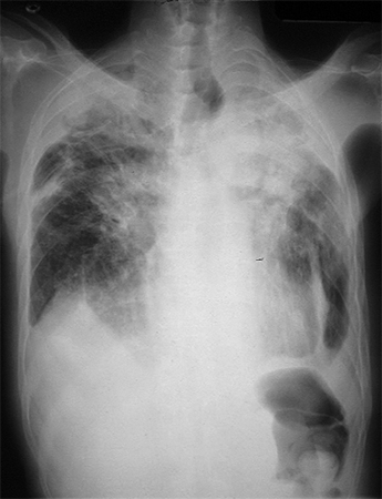
Result
silicosis and coal workers' pneumoconiosis: progressive upper zone noncalcified, small, rounded opacities, "egg shell calcification" specific for silicosis; chronic beryllium disease: progressive upper zone linear interstitial fibrosis
spirometry
Test
Nonspecific test. Useful to determine degree of impairment and pharmacologic treatment.[32]
Indicated in all patients with radiographic changes; significant silica, coal, or beryllium exposure; or shortness of breath.
Restrictive pattern: reduced FVC, normal FEV1/FVC ratio, reduced slow vital capacity, reduced total lung capacity (TLC), reduced lung diffusion capacity testing (DLCO).
Obstructive changes: reduced FEV1, reduced FEV1/FVC ratio, increased residual volume/TLC ratio, reduced DLCO.
Mixed changes: combination of restrictive and obstructive changes.
The risk of obstructive changes is increased in patients who have a history of mineral dust exposure and cigarette smoking.[26][34][35]
Reduced DLCO is the most sensitive pulmonary function change.
The American Thoracic Society and European Respiratory Society have produced joint guidelines on best practice for spirometry. ATS/ERS: standardization of spirometry 2019 update Opens in new window
Result
may be normal or demonstrate restrictive changes; may show obstructive or mixed pattern
beryllium lymphocyte proliferation test (BeLPT)
Test
Essential component of the diagnosis of chronic beryllium disease in the US.[13]
Sensitive test that identifies individuals sensitized to beryllium.
Typically performed on a blood sample first, and confirmed with a repeat test.
Bronchoscopic lavage fluid is subsequently tested for BeLPT and may be positive when the blood test is negative.
The occurrence of a positive BeLPT without lung granulomas on biopsy is an indication of sensitization to beryllium and absence of chronic beryllium disease.
Result
if sensitized to beryllium: positive response
Tests to consider
bronchoscopic biopsy and/or lavage
Test
A routine part of the diagnostic workup for chronic beryllium disease.
The occurrence of a positive beryllium lymphocyte proliferation test (BeLPT) without granulomas is an indication of sensitization to beryllium and absence of chronic beryllium disease.
Bronchoscopic biopsy may occasionally be performed in people suspected to have pneumoconiosis related to silica or coal exposure. However, generally provides insufficient tissue to rule out silicosis or coal workers' pneumoconiosis. In these patients it is limited to evaluation for cancer or other clinical conditions if suspected.
Result
chronic beryllium disease: granulomas present
high-resolution CT (HRCT) scan chest
Test
HRCT scan of the chest is more sensitive than chest x-ray in identifying interstitial fibrosis and therefore is useful when a patient has symptoms or abnormalities of pulmonary testing but their chest x-ray is normal.[29][41] It is also more sensitive in detecting when the patient has progressed from simple silicosis or coal workers' pneumoconiosis to progressive massive fibrosis and may be used to further characterize the extent of parenchymal disease following a chest x-ray.[35]
Result
upper zone interstitial fibrosis; progressively involves the entire lung
oxygen saturation
Test
Oxygen saturation may help to further define the degree of impairment.
Oxygen saturation at rest and after exercise is also useful to determine if a patient needs ongoing oxygen therapy and is indicated in individuals with significant changes on pulmonary function testing.
Result
may be reduced
arterial blood gas (ABG)
Test
Rarely performed. May further define the degree of impairment.
Result
may be a reduced pO₂; in presence of COPD, may be an elevated pCO₂
lung biopsy
Test
Rarely needed for diagnosis. Its use should be limited to when cancer is suspected, or when there is an absence of a known history of exposure to mineral dust.
Silicosis: polarized light is used to identify birefringent silica particles; silicotic nodules present.
Progressive massive fibrosis in silicosis: a mass of dense, hyalinized connective tissue present.
Acute silicosis: alveolar filling with proteinaceous material consisting largely of phospholipids or surfactants that stain with PAS reagent.
Coal workers' pneumoconiosis: focal and discrete changes related to respiratory bronchioles where phagocytosed particles are aggregated; a small amount of reticulin formation. Concomitant focal emphysema present.
Coal workers' pneumoconiosis with progressive massive fibrosis: intense carbon pigmentation, dense fibrosis, endarteritis obliterans of the blood vessels, and extensive areas of necrosis.
Chronic beryllium disease: granulomas with a center composed of epithelioid-like cells. Granulomas caused by sarcoidosis have a similar histologic appearance.
Result
variable findings
test for tuberculosis (TB)
Test
Patients with silicosis should be tested for TB.[37] The risk of active TB is even greater in those mining populations with a high prevalence of HIV infection.[38] See Pulmonary tuberculosis (Diagnostic approach).
Result
if TB present: positive
Use of this content is subject to our disclaimer