Images and videos
Images


Pneumoconioses
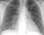
CXR of chronic beryllium disease
From the personal collection of Kenneth D. Rosenman, Michigan State University
See this image in context in the following section/s:
Pneumoconioses
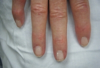
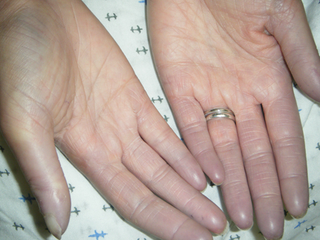
Hands demonstrating Raynaud phenomenon
From the collection of Maureen D. Mayes, University of Texas
See this image in context in the following section/s:

Pneumoconioses
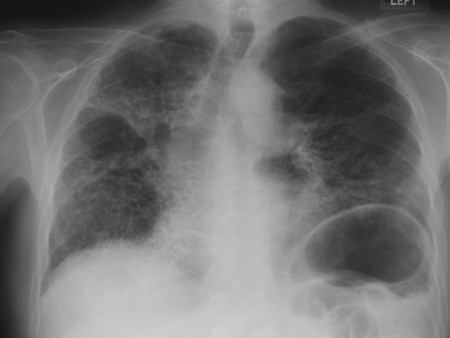
Sarcoidosis: CXR demonstrating bilateral hilar lymphadenopathy plus pulmonary infiltrates
Adapted from BMJ (2009); used with permission
See this image in context in the following section/s:
Pneumoconioses
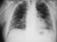
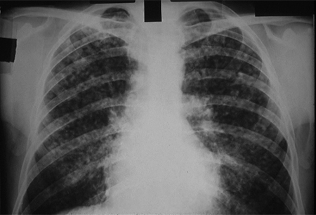
CXR showing changes consistent with simple silicosis or coal workers' pneumoconiosis
From the personal collection of Kenneth D. Rosenman, Michigan State University
See this image in context in the following section/s:

Pneumoconioses
Fingers demonstrating sclerodactyly with finger curling, shiny skin at the fingers, and telangiectasias
From the collection of Maureen D. Mayes, University of Texas
See this image in context in the following section/s:

Pneumoconioses
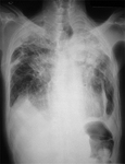
CXR of progressive massive fibrosis due to silica or coal exposure
From the personal collection of Kenneth D. Rosenman, Michigan State University
See this image in context in the following section/s:

Pneumoconioses
CXR of asbestosis
From the personal collection of Kenneth D. Rosenman, Michigan State University
See this image in context in the following section/s:
Pneumoconioses
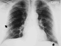
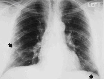
CXR demonstrating pleural thickening (indicated by arrows)
From the personal collection of Kenneth D. Rosenman, Michigan State University
See this image in context in the following section/s:
Use of this content is subject to our disclaimer