Images and videos
Images


Ventricular septal defects
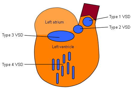
Simplified diagram of the left ventricular septum showing the anatomical locations of the ventricular septal defects
From the collection of Dr Zuhdi Lababidi
See this image in context in the following section/s:
Ventricular septal defects
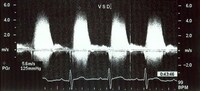
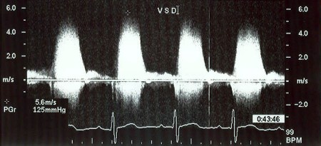
Doppler image showing spectral recording of continuous-wave Doppler showing the left-to-right gradient across the ventricular septal defect
From the collection of Dr Kul Aggarwal
See this image in context in the following section/s:
Ventricular septal defects
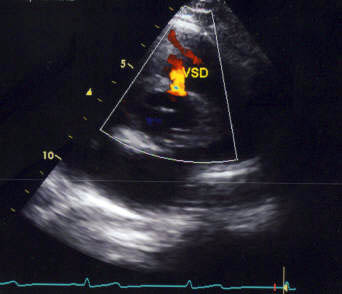
Echocardiographic image with color flow Doppler showing left-to-right shunting across a type 1 ventricular septal defect at the supracristal (or doubly committed juxta-arterial) level
From the collection of Dr Zuhdi Lababidi
See this image in context in the following section/s:
Ventricular septal defects
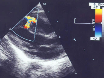
Echocardiographic image of a type 4 (muscular) ventricular septal defect with color flow Doppler showing left-to-right shunt
From the collection of Dr Kul Aggarwal
See this image in context in the following section/s:
Ventricular septal defects
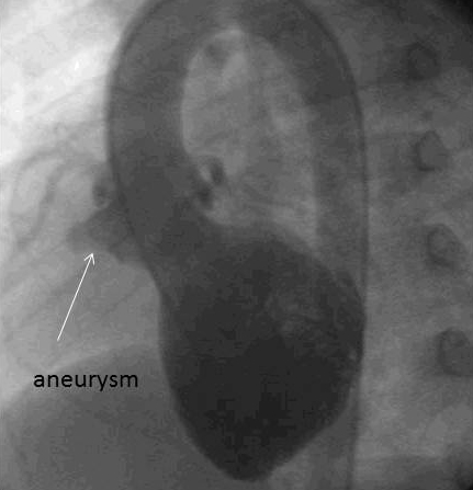
X-ray angiographic image showing an aneurysm without left-to-right shunting
From the collection of Dr Zuhdi Lababidi
See this image in context in the following section/s:
Ventricular septal defects
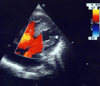
Echocardiographic image with color flow Doppler showing left-to-right shunting across a type 3 ventricular septal defect at the inlet cushion level
From the collection of Dr Zuhdi Lababidi
See this image in context in the following section/s:

Ventricular septal defects
X-ray image showing a type 4 (muscular) ventricular septal defect on left ventriculography
From the collection of Dr Zuhdi Lababidi
See this image in context in the following section/s:
Ventricular septal defects
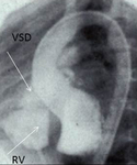
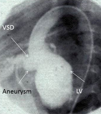
X-ray image showing a type 2 (membranous) ventricular septal defect with an aneurysm on left ventriculography
From the collection of Dr Zuhdi Lababidi
See this image in context in the following section/s:

Ventricular septal defects
Echocardiographic image showing ventricular septal defect at the inlet cushion level
From the collection of Dr Zuhdi Lababidi
See this image in context in the following section/s:
Ventricular septal defects
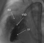
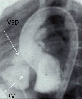
X-ray image showing a type 2 (membranous) ventricular septal defect on left ventriculography
From the collection of Dr Zuhdi Lababidi
See this image in context in the following section/s:
Use of this content is subject to our disclaimer