History and exam
Key diagnostic factors
common
shortness of breath
Likely the most common complaint, shortness of breath is a late sign. It is constant, occurring at rest or on exertion, occasionally positional with labored breathing in the recumbent position and not responsive to bronchodilators. The degree of shortness of breath does not necessarily correlate with the degree of obstruction. The diameter of the tracheal lumen must be <8 mm for dyspnea on exertion to develop and <5 mm for dyspnea at rest.[2][60][63] Individuals with pre-existing lung disease (e.g., COPD) may become symptomatic with a lesser degree of obstruction.
cough
Normally chronic, persistent, and dry, but may present acutely in foreign-body aspiration or be productive of purulent sputum in post-obstructive pneumonia.
hemoptysis
Common, especially in tracheal lesions, and may be massive, although most studies report mild to moderate hemoptysis. Squamous cell carcinoma, primary airway tumors (carcinoid tumors and adenoid cystic carcinoma), endobronchial metastases, infections (e.g., tuberculosis), inflammatory diseases (e.g., granulomatosis with polyangiitis [formerly known as Wegener granulomatosis]), and benign airway tumors (e.g., hamartomas) frequently cause hemoptysis. Chronic bronchitis frequently presents with blood-streaked purulent secretions, which may be misleading in the diagnosis of CAO.[65]
wheeze
May be inspiratory or expiratory. The location of wheeze does not always conform to the site of the airflow obstruction, and it may be heard over the trachea or lung fields.[66] Unilateral wheeze suggests obstruction distal to the carina. Wheeze may also be positional and unresponsive to bronchodilators.[2][9]
stridor
Stridor develops when the airway diameter is <5 mm and represents severe subglottic or tracheal stenosis.[66] Inspiratory stridor suggests extrathoracic airway obstruction at or above the vocal cords and is best heard over the neck,[15] while expiratory stridor may be due to an intrathoracic obstruction.[60] Biphasic stridor is present in subglottic or tracheal stenosis.[15] Maneuvers that increase airflow such as hyperventilation may accentuate the stridor, and neck flexion may change its intensity.[33]
Other diagnostic factors
uncommon
hoarseness
Hoarseness is associated with upper airway malignancies. It may also be part of the presentation of tracheal stenosis secondary to endotracheal intubation, along with dyspnea and cough.
orthopnea
Some patients may complain of positional dyspnea, with difficulty grasping air in the recumbent position due to structural compression by large intrathoracic tumors.
dysphagia
May be present in patients with large airway malignancies causing esophageal compression, or esophageal malignancies with endobronchial invasion.
chest pain
May be substernal, or located in any other thoracic site.
anxiety
In severe CAO presentations, extreme anxiety may be seen and may herald an impending respiratory arrest.
tachypnea
In severe CAO presentations, tachypnea may be seen and may herald an impending respiratory arrest.
tachycardia
In severe CAO presentations, tachycardia may be seen and may herald an impending respiratory arrest.
accessory muscle use
In severe CAO presentations, accessory muscle use as well as sternal retraction and neck extension may be seen, and may herald an impending respiratory arrest.
cyanosis
In severe CAO presentations, cyanosis may be seen and may herald an impending respiratory arrest.
crackles
Localized crackles or frank consolidation may be present in post-obstructive pneumonia.
Risk factors
strong
lung cancer
The most common cause of malignant central airway obstruction (CAO), and likely of all types of tracheobronchial obstruction, is direct extension from an adjacent tumor, most commonly bronchogenic carcinoma.[2][22] The most common cell type reported is squamous cell carcinoma, which accounts for more than half of the non-small-cell lung cancer related central airway obstructions.[29][30]
primary airway malignancy
Primary airway malignancies are relatively rare, accounting for 3% to 5% of all lung tumors. However, carcinoid tumors and adenoid cystic carcinomas are commonly centrally located.[33]
smoking
artificial airways
tracheobronchial stents
Airway stenting for malignant and nonmalignant disease has a significant complication rate ranging from 15% to 75%, including that of airway obstruction. The airway may be occluded due to mucus impaction, granulation tissue obstruction, stent migration, stent malposition, or stent fracture.[Figure caption and citation for the preceding image starts]: Chest x-ray showing right mainstem endobronchial stent occlusion with mucusFrom the collections of Jose Fernando Santacruz MD, FCCP, DAABIP and Erik Folch MD, MSc; used with permission [Citation ends].
transtracheal oxygen catheters
Although infrequently used, transtracheal oxygen catheters may cause airway obstruction by the formation of mucous balls.[16]
lung transplantation
The incidence of airway complication following lung transplantation ranges from 1.6% to 32%. Of these, bronchial stenosis, excessive exophytic tissue granulation formation, and tracheobronchomalacia may present with CAO. Bronchial stenosis is the most common airway complication seen after lung transplantation.[14][Figure caption and citation for the preceding image starts]: Post-lung transplant anastomotic bronchial stenosisFrom the collections of Jose Fernando Santacruz MD, FCCP, DAABIP and Erik Folch MD, MSc; used with permission [Citation ends].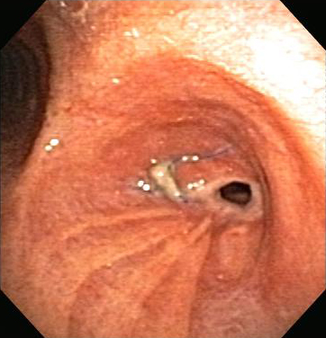
neurocognitive and neuromuscular disorders
Neurologic and neuromuscular diseases are risk factors for foreign-body aspiration in adults. Residents of nursing homes or mental health facilities, and patients with Parkinson disease, post-stroke neurocognitive sequelae, or with depressed mental status due to sedatives or alcohol are more prone to suffocation due to aspiration.[17][47]
relapsing polychondritis
Although relapsing polychondritis is a rare disease, it represents a significant risk factor, as 50% of patients with this condition will develop an airway complication. It is the most serious manifestation of the disease and predicts a poor prognosis.[55]
granulomatosis with polyangiitis (formerly known as Wegener granulomatosis)
Although a rare disease, airway involvement may be present in 15% to 55% of cases, particularly in patients <30 years of age.[10] Airway obstruction may be due to subglottic or tracheobronchial stenosis, tracheal or bronchial polyps, airway inflammation, or pseudotumors.[56] Subglottic stenosis is the most frequent airway manifestation, with a reported incidence ranging from 8.5% to 50%.[10] The incidence of bronchial stenosis is 13%.[57][Figure caption and citation for the preceding image starts]: Airway stenosis secondary to granulomatosis with polyangiitis (formerly known as Wegener granulomatosis)From the collections of Jose Fernando Santacruz MD, FCCP, DAABIP and Erik Folch MD, MSc; used with permission [Citation ends].
tracheobronchomalacia
Expiratory central airway collapse may create a flow limitation from excessive narrowing of the trachea and mainstem bronchi during exhalation that results from tracheobronchomalacia or excessive dynamic airway collapse.[13] This type of airway obstruction is often referred to as dynamic or functional stenosis.[21]
weak
endobronchial infections
Although rarely encountered in developed countries, tuberculosis (TB) endobronchial strictures have been described in 10% to 30% of TB patients undergoing bronchoscopy in Asia, Africa, and Latin America, where the disease is common. This subset clearly reflects a group of patients with airway symptoms.[45]
Histoplasmosis may cause CAO by fibrosing mediastinitis or endobronchial granulomas.[7][45]
extrathoracic and distant malignancies
Other adjacent tumors, such as thyroid, laryngeal, and esophageal cancer, may cause CAO by direct extension or airway compression. However, these malignancies are far less common than lung cancer as a cause of CAO.
Direct distal airway metastases are relatively uncommon. The most frequent extrathoracic malignancies to directly invade the airway are breast, renal cell, colorectal, and thyroid carcinomas.[35][36]
Use of this content is subject to our disclaimer