Tests
1st tests to order
CRP
Test
A marker of inflammation. Lacks specificity, as it can be raised by any inflammatory process. Sensitivity is moderate.[1][2][4][24][30] CRP is preferred to ESR in the acute phase as it has increased sensitivity and specificity to detect acute inflammation.[25]
Result
typically elevated with active disease
erythrocyte sedimentation rate (ESR)
Test
A marker of inflammation, but it lacks specificity and sensitivity. Most patients with elevated ESR do not have Takayasu arteritis, and patients with active disease can have a normal ESR.[1][2][4][24][30] CRP is preferred in the acute phase as it has increased sensitivity and specificity to detect acute inflammation.[25]
Result
typically >50 mm/hour with active disease
computed tomography angiography (CTA)
Test
CTA may be performed with helical scanning and 3D reconstruction. It has high sensitivity and specificity (over 95%) for the diagnosis of Takayasu arteritis. Catheter angiogram is not recommended due to its invasive nature, unless required prior to revascularization procedures.[26][29][31][Figure caption and citation for the preceding image starts]: Computed tomography angiogram, with 3D reconstruction of the aortic arch and major vessels, showing proximal occlusion of the left subclavian artery and patent left vertebral artery distal to the occlusion (left vertebral steal syndrome)From the collection of Kenneth J. Warrington, MD [Citation ends]. [Figure caption and citation for the preceding image starts]: Computed tomography angiogram, with 3D reconstruction of the aortic arch and major vessels, showing narrowing of the left common carotid artery and left subclavian arteryFrom the collection of Kenneth J. Warrington, MD [Citation ends].
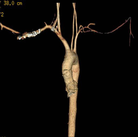 [Figure caption and citation for the preceding image starts]: Computed tomography angiogram with 3D reconstruction showing bilateral renal artery stenosisFrom the collection of Kenneth J. Warrington, MD [Citation ends].
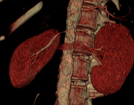
Result
segmental narrowing or occlusion, occasionally dilation, of affected vessels; aortic aneurysms may be seen; thickening of vessel walls may be seen but is of uncertain significance
magnetic resonance angiography (MRA)
Test
MRA is used to identify arterial involvement, and it may be useful in the assessment of disease activity, with vessel wall thickening and edema thought to reflect active disease.[32][Figure caption and citation for the preceding image starts]: Magnetic resonance angiogram of the aortic arch and major vessels showing occlusion of bilateral subclavian arteries; left common carotid artery has small diameter; proximal vertebral arteries are not identifiedFrom the collection of Kenneth J. Warrington, MD [Citation ends].
Result
segmental narrowing, occlusion, or dilation of involved arteries; vessel wall inflammation may be detectable
Tests to consider
Doppler ultrasound
Test
Particularly useful in the early vascular evaluation of patients with suspected Takayasu arteritis, as it is a noninvasive procedure. Abdominal ultrasound may reveal mesenteric or renal artery stenosis, and transthoracic/transesophageal studies can detect abnormalities in the upper aorta and subclavian and carotid arteries.[33]
Result
segmental narrowing, occlusion, and/or dilation of involved arteries
positron emission tomography with radiolabeled fluorodeoxyglucose (PET-FDG)
Test
Can be used to identify inflammation in the large arteries and is therefore a useful technique to establish diagnosis. PET-FDG can also be used for assessing disease activity over time.[26][27]
PET-FDG performs comparably to CTA for the detection of aortitis.[26][34]
Result
may show increased uptake in actively inflamed arterial segments
Use of this content is subject to our disclaimer