Images and videos
Images
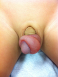


Anatomic penile abnormalities
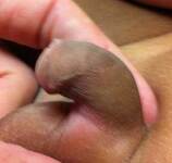
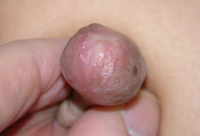
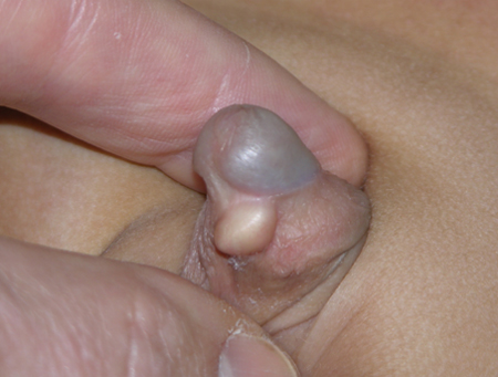
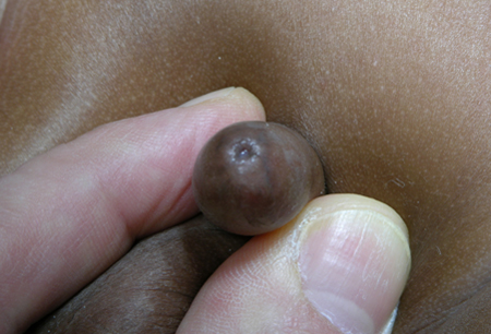
Large keratin pearl
From the collection of Warren T. Snodgrass, MD
See this image in context in the following section/s:

Anatomic penile abnormalities
Congenital penile curvature (congenital chordee)
From the collection of Nicol Corbin Bush, MD
See this image in context in the following section/s:

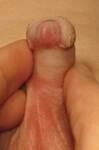
Anatomic penile abnormalities
Physiologic phimosis
From the collection of Nicol Corbin Bush, MD
See this image in context in the following section/s:
Anatomic penile abnormalities
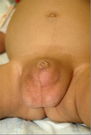
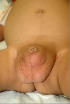
Congenital buried penis
From the collection of Nicol Corbin Bush, MD
See this image in context in the following section/s:

Anatomic penile abnormalities
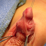
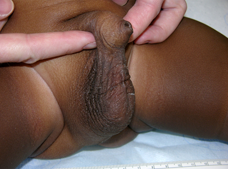
Paraphimosis
From the collection of Nicol Corbin Bush, MD
See this image in context in the following section/s:
Anatomic penile abnormalities
Balanitis xerotica obliterans (lichen sclerosis)
From the collection of Warren T. Snodgrass, MD
See this image in context in the following section/s:
Anatomic penile abnormalities
Congenital buried penis
From the collection of Warren T. Snodgrass, MD
See this image in context in the following section/s:
Anatomic penile abnormalities
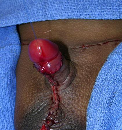
Repaired congenital buried penis
From the collection of Nicol Corbin Bush, MD
See this image in context in the following section/s:

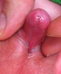
Anatomic penile abnormalities
Early paraphimosis
From the collection of Nicol Corbin Bush, MD
See this image in context in the following section/s:

Anatomic penile abnormalities
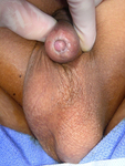
Pathologic phimosis in a patient with balanitis xerotic obliterans
From the collection of Nicol Corbin Bush, MD
See this image in context in the following section/s:
Anatomic penile abnormalities
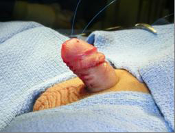
Repaired congenital buried penis (with bilateral hernia repair and scrotoplasty)
From the collection of Warren T. Snodgrass, MD
See this image in context in the following section/s:

Anatomic penile abnormalities
Infant with distal hypospadias. The urethral meatus is located in the glans or distal shaft and prepuce is typically incomplete
From the collection of Nicol Corbin Bush, MD
See this image in context in the following section/s:

Anatomic penile abnormalities
Infant with proximal hypospadias. The urethral meatus is located from the perineum to the proximal shaft
From the collection of Nicol Corbin Bush, MD
See this image in context in the following section/s:

Anatomic penile abnormalities
Congenital buried penis: abundant inner preputial skin with paucity of shaft skin
From the collection of Nicol Corbin Bush, MD
See this image in context in the following section/s:
Anatomic penile abnormalities
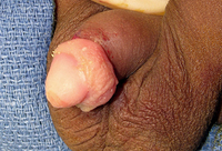
Pathologic phimosis with cicatrix
From the collection of Warren T. Snodgrass, MD
See this image in context in the following section/s:
Anatomic penile abnormalities
Congenital buried penis
From the collection of Nicol Corbin Bush, MD
See this image in context in the following section/s:

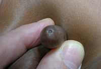
Anatomic penile abnormalities
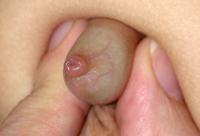
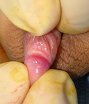
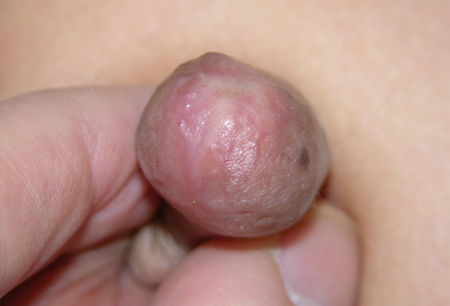
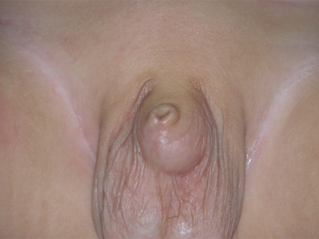
Small keratin pearls
From the collection of Nicol Corbin Bush, MD
See this image in context in the following section/s:
Use of this content is subject to our disclaimer