History and exam
Key diagnostic factors
common
newborn and toddler age
Congenital (physiologic) phimosis is expected in children <3 years of age, and may persist normally until puberty. It is present in most uncircumcised newborn males, and decreases with age (such that only about 1% of 16- to 17-year-old males are affected).[7]
Hypospadias and congenital penile curvature and/or torsion are typically diagnosed in the newborn period. Megameatus with intact prepuce (a mild variant of distal hypospadias) is diagnosed after newborn circumcision or at an older age once the foreskin is retracted.
abnormal location of urethra
Indicates hypospadias. Severity can range from the distal form (i.e., the urethral meatus is located on the glans or distal shaft) to the less common proximal form (i.e., the urethral meatus is located from the perineum to the proximal shaft).
Incomplete prepuce is a frequent finding with both proximal and distal hypospadias, though it may be intact in some cases of distal hypospadias. Penile curvature is also common, particularly in patients with proximal hypospadias; patients with proximal hypospadias may also have a cleft scrotum.
Megameatus with intact prepuce (a mild variant of distal hypospadias) can occur with an intact prepuce, and is only noted at the time of circumcision or retraction of the prepuce at an older age.[Figure caption and citation for the preceding image starts]: Infant with distal hypospadias. The urethral meatus is located in the glans or distal shaft and prepuce is typically incompleteFrom the collection of Nicol Corbin Bush, MD [Citation ends].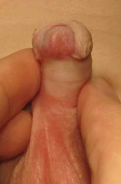 [Figure caption and citation for the preceding image starts]: Infant with proximal hypospadias. The urethral meatus is located from the perineum to the proximal shaftFrom the collection of Nicol Corbin Bush, MD [Citation ends].
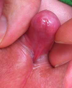
incomplete prepuce
Hypospadias is typically diagnosed at birth when the hallmark incomplete prepuce is visualized.
penile curvature and/or torsion
Congenital penile curvature and/or torsion are typically noted in the newborn period when the infant has a spontaneous erection, often with voiding. A forward or sideways penile bend is seen, sometimes with penoscrotal webbing and/or deficient ventral shaft skin. Less commonly, dorsal curvature is seen.
Penile curvature may occur in conjunction with hypospadias, or as an isolated finding. The position of the meatus should be evaluated in these patients.
Congenital penile curvature >30 degrees is considered clinically significant.[11][Figure caption and citation for the preceding image starts]: Congenital penile curvature (congenital chordee)From the collection of Nicol Corbin Bush, MD [Citation ends].
recent genital exam or procedure
history of short or small penis
The most common presenting symptom of concealed penis is a complaint from the referring provider, patient, or parent of a ''short penis.''
penile pain and swelling
Paraphimosis is almost always accompanied by penile swelling (distal to the retracted foreskin) and pain (typically the presenting symptom).
foreskin adherent to glans
Congenital (physiologic) phimosis is expected in children younger than 3 years of age, and may persist normally until puberty.
penile adhesions and smegma
Glanular adhesions between the glans and the prepuce, and a whitish collection of desquamated cells called smegma, may be visible between the glans and the prepuce.
penile cicatrix
The foreskin is characterized in severe phimosis by a constrictive, sclerotic, white ring (cicatrix) at the tip.
Boys who have recurrent episodes of balanitis or balanoposthitis are at risk of developing cicatrix, contributing to pathologic phimosis.[35]
penile glans edema
Paraphimosis is accompanied by edema of the glans with a retracted foreskin proximal to the glans penis.
prominent prepubic fat pad
Concealed penis is diagnosed when a penis is of normal length but is buried under surrounding tissues. Typically seen in neonates or overweight prepubertal boys.
presence of hernia or hydrocele
Distortion of normal anatomy leading to secondary buried penis occurs in the presence of hernia or hydrocele.
Other diagnostic factors
common
forced retraction of foreskin
dyspareunia
Pain with erections and difficulty with sexual intercourse are common complaints among adults with acquired phimosis.
uncommon
recent penile trauma
history of balanitis or balanoposthitis
Boys who have recurrent episodes of balanitis or balanoposthitis are at risk of developing scarred preputial orifices, contributing to pathologic phimosis.[35]
urinary obstruction or retention
Scarred foreskin may make urination difficult. Ballooning foreskin, postmicturition dribbling of urine, stranguria (straining to void), and even hematuria may be present.
Some patients may delay or avoid voiding owing to discomfort.
necrosis of penile skin
Necrosis of penile shaft skin may rarely occur in paraphimosis, typically after prolonged retraction of the foreskin.
discoloration of glans
Penile necrosis may occur after prolonged untreated paraphimosis.
Should be suspected with a firm, discolored glans.[41]
[Figure caption and citation for the preceding image starts]: Physiologic phimosisFrom the collection of Nicol Corbin Bush, MD [Citation ends]. [Figure caption and citation for the preceding image starts]: Pathologic phimosis with cicatrixFrom the collection of Warren T. Snodgrass, MD [Citation ends].
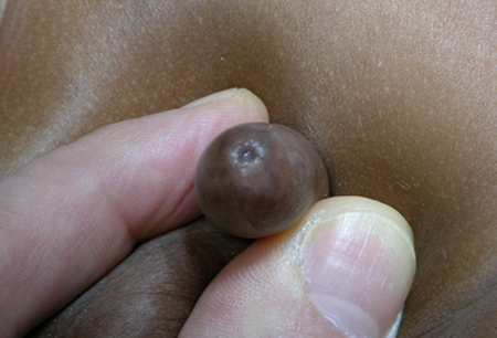 [Figure caption and citation for the preceding image starts]: Early paraphimosisFrom the collection of Nicol Corbin Bush, MD [Citation ends].
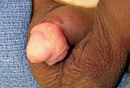 [Figure caption and citation for the preceding image starts]: Pathologic phimosis in a patient with balanitis xerotic obliteransFrom the collection of Nicol Corbin Bush, MD [Citation ends].
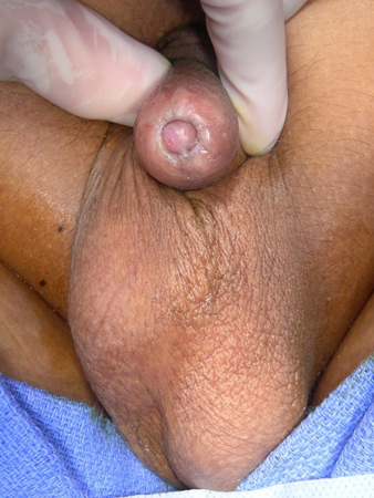 [Figure caption and citation for the preceding image starts]: ParaphimosisFrom the collection of Nicol Corbin Bush, MD [Citation ends].
penile length discrepancy
If the stretched penile length is 2.5 or more standard deviations below the mean for age, micropenis is diagnosed and endocrine evaluation is appropriate.[45]
history of urinary tract infection
As hygiene becomes an issue in cases of acquired concealed penis, the patient may present with a history of frequent skin or urine infections (though care should be taken to review laboratory data to ensure that infections are truly present, and if so, at what site).[4]
history of pelvic or genitourinary surgery
Acquired concealed penis may be caused by disruption of the suspensory tissues and tissue planes during other surgery in the pelvic or inguinal region.[36]
erectile dysfunction
Difficulty with sexual intercourse may occur among adults with concealed penis.
Risk factors
strong
uncircumcised penis (paraphimosis)
Paraphimosis occurs when the foreskin is retracted; therefore, only uncircumcised (or partially circumcised) males are at risk.[Figure caption and citation for the preceding image starts]: ParaphimosisFrom the collection of Nicol Corbin Bush, MD [Citation ends].[Figure caption and citation for the preceding image starts]: Early paraphimosisFrom the collection of Nicol Corbin Bush, MD [Citation ends].
indwelling urinary catheter (paraphimosis)
parental unawareness (phimosis)
Parents who attempt to forcibly retract phimotic foreskin may not be able to replace the foreskin afterward.
balanitis xerotica obliterans (phimosis)
Also known as lichen sclerosis.
Found on histologic exam of prepuce in 40% of boys between 2 and 16 years of age (mean 6.8 years) with acquired phimosis.[32][Figure caption and citation for the preceding image starts]: Balanitis xerotica obliterans (lichen sclerosis)From the collection of Warren T. Snodgrass, MD [Citation ends].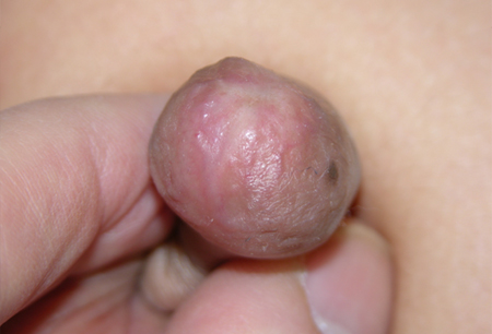
penile trauma (phimosis)
Forced retraction of congenital phimosis resulting in local trauma to the tissues, unusual masturbation practices, and repeated catheterization may result in acquired phimosis.
recurrent balanitis and balanoposthitis (phimosis)
Boys who have recurrent episodes of balanitis or balanoposthitis are at risk of developing cicatrix, contributing to pathologic phimosis.[35] In adults, concealed penis may lead to balanitis, which in turn leads to more scar tissue and worsening of the disease process.[36][Figure caption and citation for the preceding image starts]: Pathologic phimosis with cicatrixFrom the collection of Warren T. Snodgrass, MD [Citation ends].[Figure caption and citation for the preceding image starts]: Pathologic phimosis in a patient with balanitis xerotic obliteransFrom the collection of Nicol Corbin Bush, MD [Citation ends].
gestational diabetes, maternal obesity, maternal hypertension, older maternal age (hypospadias)
family history (hypospadias, congenital penile curvature and/or torsion)
weak
circumcision (trapped penis)
Trapped penis may be seen in patients who have been circumcised before realizing they have some degree of buried penis.
Simple circumcision worsens the condition in many cases, resulting in a penis that appears even more buried and which may have insufficient skin for repair. This may lead to a need for skin graft.[41] Circumcision is best performed surgically (rather than as an office procedure) in patients found to have a penis concealed within the prepubic fat.
penile and lower abdominal scarring (buried penis)
Penile or low abdominal surgery, or trauma to this area, may affect the fascial attachments of the penis, with scarring and tethering of the phallus.
hernia or hydrocele (buried penis)
Distortion of normal anatomy leading to secondary buried penis occurs in the presence of hernia or hydrocele. The condition resolves with resolution or repair of the hernia or hydrocele.
Use of this content is subject to our disclaimer