Approach
Because no single symptom, exam finding, lab test, or radiographic pathology confirms the diagnosis of adhesive capsulitis, it is often viewed as a diagnosis of exclusion. It is essential to rule out other pathology, such as acute or chronic posterior dislocation, massive rotator cuff tears, or glenohumeral arthritis prior to assuming a diagnosis of adhesive capsulitis.
History
Patient sex, age, hand dominance, and occupation are important demographic factors, not only for diagnosis but also for eventual management.
Information regarding duration of pain (if any), location of pain, timing of pain (including night pain or difficulties sleeping due to pain), exacerbating activities (including sleeping on the affected side), limitations of work or daily activities, and any concomitant pain, numbness, or paresthesias must be obtained. Pain in other areas around the shoulder joint, or neurologic symptoms, are suggestive of another diagnosis.
Patients will often report insidious onset of pain followed by decreased range of shoulder motion, even after resolution of pain. Frequently, this limitation of motion has hindered the patient's ability to work or to perform activities of daily living.
Patients may present in any one of the 4 classic stages of adhesive capsulitis:
Stage 1: patients may complain of lateral shoulder pain, especially at night, but may only have small limitations in motion.
Stage 2: range of motion decreases to a greater extent and pain becomes more severe.
Stage 3: pain is limited only to extremes of range of motion but motion is now severely limited.
Stage 4: there is negligible pain but profound loss of motion.
After stage 4, stiffness gradually resolves.[28] Physical therapy and home exercises may be limited in the early stages of the disease, when pain is more severe.
In a minority of cases, the patient may have diabetes or a thyroid disorder, both factors that may contribute to the development of secondary adhesive capsulitis.
A history of injury and/or previous surgery to the affected shoulder, and the duration of immobilization, are important to establish.
Physical examination
Patients with adhesive capsulitis will be limited in both active and passive range of motion, regardless of whether or not they have pain. Therefore, accurate measurements of active and passive range of motion are needed.
Patients with adhesive capsulitis will have limited motion of the glenohumeral joint, and may attempt to compensate for this by moving the arm via scapulothoracic motion. The examination is performed in the supine position with the scapula locked in position by gravity against the table. This allows the clinician to isolate glenohumeral motion. The range of motion testing is performed with flexion, abduction, and then abducted internal and external rotation, with the humerus abducted to 90°. (Normal abducted external rotation is 90°; normal abducted internal rotation is approximately 75°.) The glenohumeral joint will have restricted range of motion in adhesive capsulitis. The differences in motion between the affected shoulder and the contralateral shoulder, as well as the relative loss of external and internal rotation on the affected side, are important to note.
There may be pain at the limitations of motion, which signifies capsular tightness, synovitis, and irritation.
The coracoid pain test is a highly sensitive and specific clinical exam finding for adhesive capsulitis. Pain elicited by direct pressure on the coracoid was 96% sensitive and 87% to 89% specific in one study describing the test.[29]
According to one systematic review of physical examination tests for the shoulder, the shoulder shrug test (in which an inability to abduct the arm to 90° in the plane of the body and to hold that position briefly is considered positive) is 95% sensitive and 50% specific for adhesive capsulitis. However, it is important to note that the authors denote this test as a nonspecific sign for other shoulder pathologies, such as glenohumeral osteoarthritis or massive rotator cuff tears.[30]
Provocative examination maneuvers for labral and rotator cuff pathology, as well as cervical spine pathology, can be used to exclude other potential causes for decreased shoulder range of motion.[Figure caption and citation for the preceding image starts]: Spurling maneuverFrom the private collection of Matthew T. Provencher, MD, CDR MC USN and Lance E. LeClere, MD, LCDR MC USN; used with permission [Citation ends]. [Figure caption and citation for the preceding image starts]: O'Brien testFrom the private collection of Matthew T. Provencher, MD, CDR MC USN and Lance E. LeClere, MD, LCDR MC USN; used with permission [Citation ends].
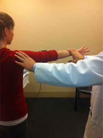 [Figure caption and citation for the preceding image starts]: Yergason testFrom the private collection of Matthew T. Provencher, MD, CDR MC USN and Lance E. LeClere, MD, LCDR MC USN; used with permission [Citation ends].
 [Figure caption and citation for the preceding image starts]: Apprehension testFrom the private collection of Matthew T. Provencher, MD, CDR MC USN and Lance E. LeClere, MD, LCDR MC USN; used with permission [Citation ends].
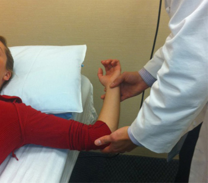 [Figure caption and citation for the preceding image starts]: Kim testFrom the private collection of Matthew T. Provencher, MD, CDR MC USN and Lance E. LeClere, MD, LCDR MC USN; used with permission [Citation ends].
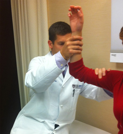
[Figure caption and citation for the preceding image starts]: Examples of provocative examination maneuvers used to exclude labral, rotator cuff, and cervical spine pathologyFrom Matthew T. Provencher, MD, CDR MC USN and Lance E. LeClere, MD, LCDR MC USN; used with permission [Citation ends].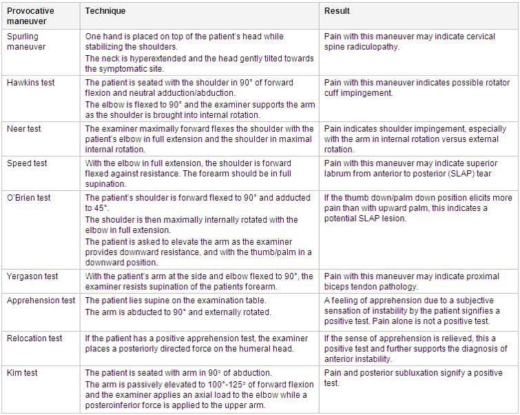
Imaging
No single imaging study is diagnostic. Plain radiographs (anteroposterior, lateral, and axillary views of the glenohumeral joint) are the preferred initial test to rule out other potential shoulder pathologies.[31]
In certain instances where clinical examination suggests adhesive capsulitis, symptoms of impingement, or painful palpable mass, ultrasound (US) may be considered as an initial imaging study. US has the advantage of allowing provocative maneuvers to assess impingement and can help exclude a rotator cuff tear.[31][32][33]
Advanced imaging with magnetic resonance imaging (MRI)/magnetic resonance (MR) arthrogram may aid in confirming the diagnosis of adhesive capsulitis by demonstrating a decreased capsular volume and size, and changes in rotator interval dimensions, as well as capsular thickening of the affected joint.[31][34][35]
The MRI/MR arthrogram is also useful to demonstrate a lack of concomitant shoulder pathology, such as labral or rotator cuff tears, in patients with suspected adhesive capsulitis. Computed tomography (CT) arthrogram, if easier to obtain, permits assessment of capsular volume and size.
However, MR arthrogram and CT arthrogram are not routinely recommended when evaluating for adhesive capsulitis due to conflicting results of efficacy studies.[31][36][37]
Use of this content is subject to our disclaimer