Images and videos
Images
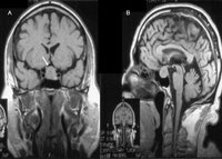

Pituitary adenoma
Precontrast coronal (A) and sagittal (B) MR images of a patient with a pituitary macroadenoma (arrow, A). The pituitary mass extends toward the optic chiasm with some pressure effect
From the collection of Dr Amir Hamrahian
See this image in context in the following section/s:

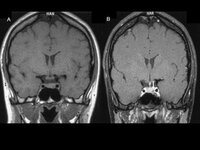
Pituitary adenoma
Precontrast (A) and postcontrast (B) coronal MR images of a patient with a small pituitary microadenoma. The pituitary lesion enhances less than the normal pituitary gland following gadolinium and appears hypodense compared with the normal pituitary gland (arrow, B)
From the collection of Dr Amir Hamrahian
See this image in context in the following section/s:
Use of this content is subject to our disclaimer