History and exam
Key diagnostic factors
common
skin patches, plaques, or tumors
It is possible to have one or all three types of lesions.
Many patients will have had the skin lesions for a number of years and been treated for other skin disorders such as psoriasis, eczema, or an allergic reaction.
Patches are usually flat, and may be scaly. Plaques are thicker, raised lesions, while tumors are raised lesions that may or may not ulcerate.
Any body surface can be affected, including the palms and the soles of the feet, but in most early presentations of mycosis fungoides (MF) the lesions are limited to the chest, abdomen, pelvis, and back.[37][38] Facial involvement is uncommon in the early stages, except in the folliculotropic variant of MF.
Patients with MF presenting in the later stages may have generalized skin lesions.[Figure caption and citation for the preceding image starts]: Cutaneous T-cell lymphoma: early patch diseaseFrom the collection of the Christie NHS Foundation Trust, Manchester, UK; used with permission [Citation ends].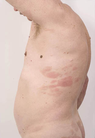 [Figure caption and citation for the preceding image starts]: Cutaneous T-cell lymphoma: extensive patch diseaseFrom the collection of the Christie NHS Foundation Trust, Manchester, UK; used with permission [Citation ends].
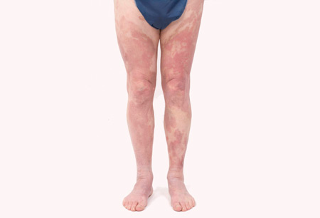 [Figure caption and citation for the preceding image starts]: Tumor plus ulceration in a patient with folliculotropic mycosis fungoidesFrom the collection of the Christie NHS Foundation Trust, Manchester, UK; used with permission [Citation ends].
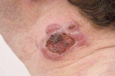
poikiloderma
Describes clinical features of atrophy, telangiectasia, and pigmentation.
uncommon
erythroderma
>90% of skin surface involved, with diffuse erythematous changes; seen in erythrodermic CTCL and Sézary syndrome. [Figure caption and citation for the preceding image starts]: ErythrodermaFrom the collection of the Christie NHS Foundation Trust, Manchester, UK; used with permission [Citation ends].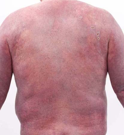
Other diagnostic factors
common
pruritus
Pruritus is one of the most frequent and debilitating symptoms of CTCL, especially for patients with Sézary syndrome. One retrospective analysis demonstrated that not only is pruritus present in over 50% of patients, but the severity of the pruritus increased with more advanced disease.[33]
hypopigmented/hyperpigmented skin lesions
Atypical presentation of mycosis fungoides. Particularly seen in childhood and adolescence and in darker skin.
unilesional acral site involvement
Atypical presentation of mycosis fungoides (Worringer-Kollop disease).
uncommon
lymphadenopathy
May be evident in patients presenting in later stages. Lymph nodes may be palpable where they drain affected skin, such as in axillary and inguinal regions.
constitutional symptoms
Patients with erythrodermic disease may experience malaise, weight loss, and chills.
Aggressive manifestations of CTCL may present with systemic symptoms of fever, night sweats, and weight loss.
palmar-plantar keratoderma
Diffuse thickening and scaling of palms and soles; seen in Sézary syndrome.
alopecia
Can occur in follicular mycosis fungoides or Sézary syndrome.
leonine facies
May be seen in Sézary syndrome.
onychodystrophy
Nail malformations; may be seen in Sézary syndrome.
hepatomegaly
May be evident at time of initial presentation in Sézary syndrome.
ectropion
Eversion of lower eyelids, due to erythroderma and edema; seen in Sézary syndrome.
bullous, granulomatous, ichthyosiform, and purpuric lesions
Rare findings associated with mycosis fungoides.
Risk factors
strong
age >50 years
male sex
Men have a higher incidence rate of cutaneous T-cell lymphomas than women. A global male to female ratio of 1.7:1 has been reported.[3]
black ethnicity (MF); white ethnicity (SS)
Overall, the incidence of cutaneous T-cell lymphomas has been shown to be higher among African-Americans, compared with white Americans and other racial groups.[7][8] Mycosis fungoides is twice as common in black Americans as it is in white Americans.[9] However, Sézary syndrome appears to have a higher incidence among white Americans.[10]
weak
exposure to infectious agents
Infectious agents investigated as potential causative agents in cutaneous T-cell lymphomas (CTCL) include Staphylococcus aureus, retro-viruses, herpes-viruses, polyomavirus, and human T-cell lymphotropic virus 1.[19][20] Studies have failed to find a consistent association between these infectious agents and CTCL. However, there is evidence to suggest that the most frequently clonally expanded T-cell receptor beta variable (TRBV) gene in SS is TRBV20-1, which is associated with the recognition of S aureus.[13] S aureus has been demonstrated to act as a superantigen and stimulate the proliferation of malignant T-cells in patients with mycosis fungoides/Sézary syndrome.[21][22]
ultraviolet light exposure
One study detected the ultraviolet marker signature 7 mutations in the blood of patients with Sézary syndrome, which may imply a role of UV exposure in cutaneous T-cell lymphomas pathogenesis.[23]
Use of this content is subject to our disclaimer