Cutaneous T-cell lymphomas (CTCL) is a heterogeneous group of malignancies, and presentation can be diverse. Establishing the correct diagnosis is often difficult and requires expertise located in specialist centers. Diagnosis is based on a combination of clinical, histologic, immunophenotypic, and genetic data.[5]Gilson D, Whittaker SJ, Child FJ, et al. Joint British Association of Dermatologists and UK Cutaneous Lymphoma Group guidelines for the management of primary cutaneous T-cell lymphomas. Br J Dermatol. 2019 Mar;180(3):496-526.
https://onlinelibrary.wiley.com/doi/10.1111/bjd.17240
http://www.ncbi.nlm.nih.gov/pubmed/30561020?tool=bestpractice.com
[31]Willemze R, Hodak E, Zinzani P, et al; ESMO Guidelines Working Group. Primary cutaneous lymphomas: ESMO clinical practice guidelines for diagnosis, treatment and follow-up. Ann Oncol. 2018 Oct 1;29(4 suppl):iv30-40.
https://www.annalsofoncology.org/article/S0923-7534(19)31693-X/fulltext
http://www.ncbi.nlm.nih.gov/pubmed/29878045?tool=bestpractice.com
[32]National Comprehensive Cancer Network. NCCN clinical practice guidelines in oncology: primary cutaneous lymphoma [internet publication].
https://www.nccn.org/guidelines/category_1
History and physical exam
Patients with suspected primary CTCL are usually older. The incidence rates for mycosis fungoides (MF)/Sézary syndrome (SS), the two most common forms of CTCL, peak in the 50- to 74-year age group, with men more frequently affected than women.[3]Dummer R, Vermeer MH, Scarisbrick JJ, et al. Cutaneous T cell lymphoma. Nat Rev Dis Primers. 2021 Aug 26;7(1):61.
http://www.ncbi.nlm.nih.gov/pubmed/34446710?tool=bestpractice.com
[4]Bradford PT, Devesa SS, Anderson WF, et al. Cutaneous lymphoma incidence patterns in the United States: a population-based study of 3884 cases. Blood. 2009 May 21;113(21):5064-73.
https://www.doi.org/10.1182/blood-2008-10-184168
http://www.ncbi.nlm.nih.gov/pubmed/19279331?tool=bestpractice.com
[30]National Cancer Information Network. Registration of cutaneous T-cell lymphoma (CTCL) in England. Oct 2016 [internet publication].
http://www.ncin.org.uk/view?rid=3275
Higher incidence rates are also found in African-Americans compared with other racial groups.[3]Dummer R, Vermeer MH, Scarisbrick JJ, et al. Cutaneous T cell lymphoma. Nat Rev Dis Primers. 2021 Aug 26;7(1):61.
http://www.ncbi.nlm.nih.gov/pubmed/34446710?tool=bestpractice.com
Evidence suggests that MF is twice as common in African-American populations, while higher rates of SS are seen in white Americans.[7]Hinds GA, Heald P. Cutaneous T-cell lymphoma in skin of color. J Am Acad Dermatol. 2009 Mar;60(3):359-75; quiz 376-8.
http://www.ncbi.nlm.nih.gov/pubmed/19231637?tool=bestpractice.com
[8]Wu XC, Andrews P, Chen VW, et al. Incidence of extranodal non-Hodgkin lymphomas among whites, blacks, and Asians/Pacific Islanders in the United States: anatomic site and histology differences. Cancer Epidemiol. 2009 Nov;33(5):337-46.
http://www.ncbi.nlm.nih.gov/pubmed/19853554?tool=bestpractice.com
[9]Weinstock MA, Horm JW. Mycosis fungoides in the United States. Increasing incidence and descriptive epidemiology. JAMA. 1988 Jul 1;260(1):42-6.
http://www.ncbi.nlm.nih.gov/pubmed/3379722?tool=bestpractice.com
[10]Crandon S, Yancey MA. Sezary syndrome: a case study of cutaneous T-cell lymphoma. Clin J Oncol Nurs. 2009 Apr;13(2):157-9.
http://www.ncbi.nlm.nih.gov/pubmed/19349262?tool=bestpractice.com
Pruritus is one of the most frequent and debilitating symptoms of CTCL, especially for patients with SS.[2]Willemze R, Cerroni L, Kempf W, et al. The 2018 update of the WHO-EORTC classification for primary cutaneous lymphomas. Blood. 2019 Apr 18;133(16):1703-14.
https://www.ncbi.nlm.nih.gov/pmc/articles/PMC6473500
http://www.ncbi.nlm.nih.gov/pubmed/30635287?tool=bestpractice.com
One retrospective analysis demonstrated that not only is pruritus present in over 50% of patients, but the severity of the pruritus increased with more advanced disease.[33]Vij A, Duvic M. Prevalence and severity of pruritus in cutaneous T cell lymphoma. Int J Dermatol. 2012 Aug;51(8):930-4.
http://www.ncbi.nlm.nih.gov/pubmed/22788808?tool=bestpractice.com
All patients should undergo a complete skin exam assessing the percentage of body surface area (BSA) and type of skin lesion, palpation of the peripheral lymph node regions, and palpation of organomegaly/masses.[31]Willemze R, Hodak E, Zinzani P, et al; ESMO Guidelines Working Group. Primary cutaneous lymphomas: ESMO clinical practice guidelines for diagnosis, treatment and follow-up. Ann Oncol. 2018 Oct 1;29(4 suppl):iv30-40.
https://www.annalsofoncology.org/article/S0923-7534(19)31693-X/fulltext
http://www.ncbi.nlm.nih.gov/pubmed/29878045?tool=bestpractice.com
[32]National Comprehensive Cancer Network. NCCN clinical practice guidelines in oncology: primary cutaneous lymphoma [internet publication].
https://www.nccn.org/guidelines/category_1
MF and its variants are characterized by heterogeneous cutaneous manifestations including:[5]Gilson D, Whittaker SJ, Child FJ, et al. Joint British Association of Dermatologists and UK Cutaneous Lymphoma Group guidelines for the management of primary cutaneous T-cell lymphomas. Br J Dermatol. 2019 Mar;180(3):496-526.
https://onlinelibrary.wiley.com/doi/10.1111/bjd.17240
http://www.ncbi.nlm.nih.gov/pubmed/30561020?tool=bestpractice.com
[32]National Comprehensive Cancer Network. NCCN clinical practice guidelines in oncology: primary cutaneous lymphoma [internet publication].
https://www.nccn.org/guidelines/category_1
[34]Bi MY, Curry JL, Christiano AM, et al. The spectrum of hair loss in patients with mycosis fungoides and Sézary syndrome. J Am Acad Dermatol. 2011 Jan;64(1):53-63.
https://www.doi.org/10.1016/j.jaad.2009.12.056
http://www.ncbi.nlm.nih.gov/pubmed/21036417?tool=bestpractice.com
[35]Kodama K, Fink-Puches R, Massone C, et al. Papular mycosis fungoides: a new clinical variant of early mycosis fungoides. J Am Acad Dermatol. 2005 Apr;52(4):694-8.
http://www.ncbi.nlm.nih.gov/pubmed/15793526?tool=bestpractice.com
[36]Boulos S, Vaid R, Aladily TN, et al. Clinical presentation, immunopathology, and treatment of juvenile-onset mycosis fungoides: a case series of 34 patients. J Am Acad Dermatol. 2014 Dec;71(6):1117-26.
http://www.ncbi.nlm.nih.gov/pubmed/25264240?tool=bestpractice.com
Polymorphic patches or plaques
Tumors and erythroderma (associated with advanced cutaneous stage)
Folliculotropic MF (FMF) may present as folliculocentric papules, nodules, or areas of alopecia
Hypopigmented lesions
Hyperpigmentation, mostly seen in children, adolescents, and patients with darker skin
Granulomatous slack skin (redundant skin resembling cutis laxa on flexural areas)
Unilesional, pagetoid reticulosis
Peripheral adenopathy may or may not be present
Any body surface can be affected, including the palms and the soles of the feet, but in most early presentations of MF the lesions are limited to the chest, abdomen, pelvis, and back.[37]Kazakov DV, Burg G, Kempf W. Clinicopathological spectrum of mycosis fungoides. J Eur Acad Dermatol Venereol. 2004 Jul;18(4):397-415.
http://www.ncbi.nlm.nih.gov/pubmed/15196152?tool=bestpractice.com
[38]Pimpinelli N, Olsen EA, Santucci M, et al. Defining early mycosis fungoides. J Am Acad Dermatol. 2005 Dec;53(6):1053-63.
http://www.ncbi.nlm.nih.gov/pubmed/16310068?tool=bestpractice.com
Facial involvement is uncommon in the early stages, except in the folliculotropic variant of mycosis fungoides. Patients with mycosis fungoides presenting in the later stages may have generalized skin lesions. Many patients will have had the skin lesions for a number of years and been treated for other skin disorders such as psoriasis, eczema, or an allergic reaction.[Figure caption and citation for the preceding image starts]: ErythrodermaFrom the collection of the Christie NHS Foundation Trust, Manchester, UK; used with permission [Citation ends].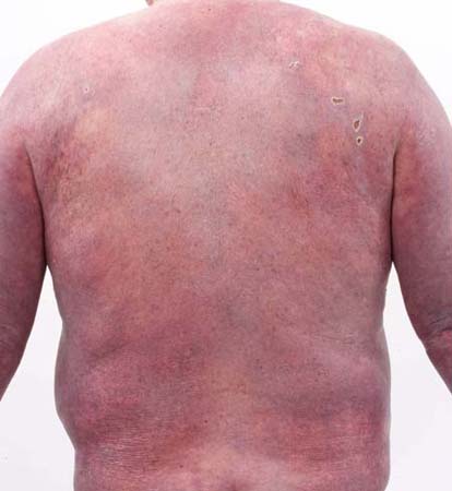 [Figure caption and citation for the preceding image starts]: Cutaneous T-cell lymphoma: early patch diseaseFrom the collection of the Christie NHS Foundation Trust, Manchester, UK; used with permission [Citation ends].
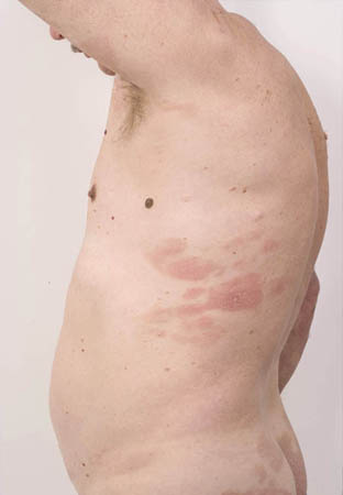 [Figure caption and citation for the preceding image starts]: Cutaneous T-cell lymphoma: extensive patch diseaseFrom the collection of the Christie NHS Foundation Trust, Manchester, UK; used with permission [Citation ends].
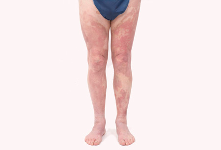 [Figure caption and citation for the preceding image starts]: Tumor plus ulceration in a patient with folliculotropic mycosis fungoidesFrom the collection of the Christie NHS Foundation Trust, Manchester, UK; used with permission [Citation ends].
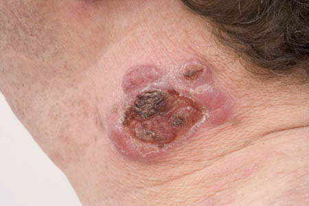
SS is a rare (<5% cases) leukemic type of CTCL, defined by:[2]Willemze R, Cerroni L, Kempf W, et al. The 2018 update of the WHO-EORTC classification for primary cutaneous lymphomas. Blood. 2019 Apr 18;133(16):1703-14.
https://www.ncbi.nlm.nih.gov/pmc/articles/PMC6473500
http://www.ncbi.nlm.nih.gov/pubmed/30635287?tool=bestpractice.com
[5]Gilson D, Whittaker SJ, Child FJ, et al. Joint British Association of Dermatologists and UK Cutaneous Lymphoma Group guidelines for the management of primary cutaneous T-cell lymphomas. Br J Dermatol. 2019 Mar;180(3):496-526.
https://onlinelibrary.wiley.com/doi/10.1111/bjd.17240
http://www.ncbi.nlm.nih.gov/pubmed/30561020?tool=bestpractice.com
[32]National Comprehensive Cancer Network. NCCN clinical practice guidelines in oncology: primary cutaneous lymphoma [internet publication].
https://www.nccn.org/guidelines/category_1
Pruritic erythroderma
Generalized lymphadenopathy
Clonally related neoplastic T cells with cerebriform nuclei (Sézary cells) in the skin, lymph nodes, and peripheral blood.
As the clinical and histologic presentation of SS may be similar to MF, demonstration of peripheral blood involvement may be crucial for the diagnosis of SS.[2]Willemze R, Cerroni L, Kempf W, et al. The 2018 update of the WHO-EORTC classification for primary cutaneous lymphomas. Blood. 2019 Apr 18;133(16):1703-14.
https://www.ncbi.nlm.nih.gov/pmc/articles/PMC6473500
http://www.ncbi.nlm.nih.gov/pubmed/30635287?tool=bestpractice.com
[5]Gilson D, Whittaker SJ, Child FJ, et al. Joint British Association of Dermatologists and UK Cutaneous Lymphoma Group guidelines for the management of primary cutaneous T-cell lymphomas. Br J Dermatol. 2019 Mar;180(3):496-526.
https://onlinelibrary.wiley.com/doi/10.1111/bjd.17240
http://www.ncbi.nlm.nih.gov/pubmed/30561020?tool=bestpractice.com
For a definitive diagnosis of SS, in addition to peripheral clonally related neoplastic T cells in skin and peripheral blood, one of the following should be present:[2]Willemze R, Cerroni L, Kempf W, et al. The 2018 update of the WHO-EORTC classification for primary cutaneous lymphomas. Blood. 2019 Apr 18;133(16):1703-14.
https://www.ncbi.nlm.nih.gov/pmc/articles/PMC6473500
http://www.ncbi.nlm.nih.gov/pubmed/30635287?tool=bestpractice.com
[5]Gilson D, Whittaker SJ, Child FJ, et al. Joint British Association of Dermatologists and UK Cutaneous Lymphoma Group guidelines for the management of primary cutaneous T-cell lymphomas. Br J Dermatol. 2019 Mar;180(3):496-526.
https://onlinelibrary.wiley.com/doi/10.1111/bjd.17240
http://www.ncbi.nlm.nih.gov/pubmed/30561020?tool=bestpractice.com
Absolute Sézary cell count of >1000/microliter
An expanded CD4+ T-cell population resulting in a CD4/CD8 ratio ≥10, CD4+/CD7- cells ≥30%, or CD4+/CD26- cells ≥40%.
Correlation of clinical features with the histopathologic findings of blood are essential for a definitive diagnosis of SS.[5]Gilson D, Whittaker SJ, Child FJ, et al. Joint British Association of Dermatologists and UK Cutaneous Lymphoma Group guidelines for the management of primary cutaneous T-cell lymphomas. Br J Dermatol. 2019 Mar;180(3):496-526.
https://onlinelibrary.wiley.com/doi/10.1111/bjd.17240
http://www.ncbi.nlm.nih.gov/pubmed/30561020?tool=bestpractice.com
[32]National Comprehensive Cancer Network. NCCN clinical practice guidelines in oncology: primary cutaneous lymphoma [internet publication].
https://www.nccn.org/guidelines/category_1
Biopsy
Biopsy of suspicious lesions is essential for all patients.[31]Willemze R, Hodak E, Zinzani P, et al; ESMO Guidelines Working Group. Primary cutaneous lymphomas: ESMO clinical practice guidelines for diagnosis, treatment and follow-up. Ann Oncol. 2018 Oct 1;29(4 suppl):iv30-40.
https://www.annalsofoncology.org/article/S0923-7534(19)31693-X/fulltext
http://www.ncbi.nlm.nih.gov/pubmed/29878045?tool=bestpractice.com
[32]National Comprehensive Cancer Network. NCCN clinical practice guidelines in oncology: primary cutaneous lymphoma [internet publication].
https://www.nccn.org/guidelines/category_1
Multiple biopsies may be necessary to capture the pathologic variability of disease. Analysis of the tumor should be done by a pathologist with expertise in the diagnosis of CTCLs.
Repeated biopsy may be necessary if the analysis of the consult material in conjunction with the clinical features is not diagnostic.[5]Gilson D, Whittaker SJ, Child FJ, et al. Joint British Association of Dermatologists and UK Cutaneous Lymphoma Group guidelines for the management of primary cutaneous T-cell lymphomas. Br J Dermatol. 2019 Mar;180(3):496-526.
https://onlinelibrary.wiley.com/doi/10.1111/bjd.17240
http://www.ncbi.nlm.nih.gov/pubmed/30561020?tool=bestpractice.com
[32]National Comprehensive Cancer Network. NCCN clinical practice guidelines in oncology: primary cutaneous lymphoma [internet publication].
https://www.nccn.org/guidelines/category_1
Immunohistochemical (IHC) panel of skin biopsy should include:[32]National Comprehensive Cancer Network. NCCN clinical practice guidelines in oncology: primary cutaneous lymphoma [internet publication].
https://www.nccn.org/guidelines/category_1
CD2, CD3, CD4, CD5, CD7, CD8, CD20, CD30
Molecular analysis to detect clonal T-cell receptor (TCR) gene rearrangements, or other assessment of clonality.
Tumor cells are usually CD3+, CD4+, and CD8-, although CD8+ variants are not uncommon. CTCL lesions often lose CD7.[5]Gilson D, Whittaker SJ, Child FJ, et al. Joint British Association of Dermatologists and UK Cutaneous Lymphoma Group guidelines for the management of primary cutaneous T-cell lymphomas. Br J Dermatol. 2019 Mar;180(3):496-526.
https://onlinelibrary.wiley.com/doi/10.1111/bjd.17240
http://www.ncbi.nlm.nih.gov/pubmed/30561020?tool=bestpractice.com
[32]National Comprehensive Cancer Network. NCCN clinical practice guidelines in oncology: primary cutaneous lymphoma [internet publication].
https://www.nccn.org/guidelines/category_1
Repeated biopsy of skin should be done if the presence of large cell transformation (histologically as greater than 25% of the tumor cells displaying large size) or folliculotropism is suspected, but not previously confirmed pathologically.[32]National Comprehensive Cancer Network. NCCN clinical practice guidelines in oncology: primary cutaneous lymphoma [internet publication].
https://www.nccn.org/guidelines/category_1
The presence of either of these variants may have important implications for selection of therapy and outcome, and should be included in pathology reports.[32]National Comprehensive Cancer Network. NCCN clinical practice guidelines in oncology: primary cutaneous lymphoma [internet publication].
https://www.nccn.org/guidelines/category_1
IHC panel of skin biopsy should include:[32]National Comprehensive Cancer Network. NCCN clinical practice guidelines in oncology: primary cutaneous lymphoma [internet publication].
https://www.nccn.org/guidelines/category_1
CD25, CD56, TIA1, granzyme B, TCRß, TCRẟ; CXCL13, inducible T-cell co-stimulator (ICOS), and PD-1
If the skin biopsy is not diagnostic, biopsy of enlarged lymph nodes or suspected extracutaneous sites may be needed.[31]Willemze R, Hodak E, Zinzani P, et al; ESMO Guidelines Working Group. Primary cutaneous lymphomas: ESMO clinical practice guidelines for diagnosis, treatment and follow-up. Ann Oncol. 2018 Oct 1;29(4 suppl):iv30-40.
https://www.annalsofoncology.org/article/S0923-7534(19)31693-X/fulltext
http://www.ncbi.nlm.nih.gov/pubmed/29878045?tool=bestpractice.com
[32]National Comprehensive Cancer Network. NCCN clinical practice guidelines in oncology: primary cutaneous lymphoma [internet publication].
https://www.nccn.org/guidelines/category_1
Excisional or incisional biopsy is the preferred method of lymph node biopsy.[32]National Comprehensive Cancer Network. NCCN clinical practice guidelines in oncology: primary cutaneous lymphoma [internet publication].
https://www.nccn.org/guidelines/category_1
A fine needle biopsy alone is not sufficient for the initial diagnosis of lymphoma. Bone marrow biopsy may be helpful in patients with unexplained hematologic abnormality. Repeated biopsy may be necessary if results are not diagnostic.[32]National Comprehensive Cancer Network. NCCN clinical practice guidelines in oncology: primary cutaneous lymphoma [internet publication].
https://www.nccn.org/guidelines/category_1
Bone marrow biopsy may be helpful in patients with unexplained hematologic abnormality.[5]Gilson D, Whittaker SJ, Child FJ, et al. Joint British Association of Dermatologists and UK Cutaneous Lymphoma Group guidelines for the management of primary cutaneous T-cell lymphomas. Br J Dermatol. 2019 Mar;180(3):496-526.
https://onlinelibrary.wiley.com/doi/10.1111/bjd.17240
http://www.ncbi.nlm.nih.gov/pubmed/30561020?tool=bestpractice.com
[32]National Comprehensive Cancer Network. NCCN clinical practice guidelines in oncology: primary cutaneous lymphoma [internet publication].
https://www.nccn.org/guidelines/category_1
Laboratory evaluation
Essential laboratory studies for all patients include:[5]Gilson D, Whittaker SJ, Child FJ, et al. Joint British Association of Dermatologists and UK Cutaneous Lymphoma Group guidelines for the management of primary cutaneous T-cell lymphomas. Br J Dermatol. 2019 Mar;180(3):496-526.
https://onlinelibrary.wiley.com/doi/10.1111/bjd.17240
http://www.ncbi.nlm.nih.gov/pubmed/30561020?tool=bestpractice.com
[31]Willemze R, Hodak E, Zinzani P, et al; ESMO Guidelines Working Group. Primary cutaneous lymphomas: ESMO clinical practice guidelines for diagnosis, treatment and follow-up. Ann Oncol. 2018 Oct 1;29(4 suppl):iv30-40.
https://www.annalsofoncology.org/article/S0923-7534(19)31693-X/fulltext
http://www.ncbi.nlm.nih.gov/pubmed/29878045?tool=bestpractice.com
[32]National Comprehensive Cancer Network. NCCN clinical practice guidelines in oncology: primary cutaneous lymphoma [internet publication].
https://www.nccn.org/guidelines/category_1
Complete blood count (CBC) with differential and determination of absolute lymphocyte count
Flow cytometric studies to assess and quantitate an expanded T-cell population with aberrant phenotype: recommended for patients with T2-4 skin classification, or any suspected extracutaneous disease including adenopathy, but optional for patients with T1 stage disease
Clonal TCR gene rearrangement in peripheral blood lymphocytes if blood involvement suspected
Comprehensive metabolic panel
Lactate dehydrogenase (LDH)
For patients with extensive skin disease where the skin biopsy is not diagnostic of advanced-stage disease, assessment of peripheral blood for Sézary cells with flow cytometry and molecular analysis is recommended.[32]National Comprehensive Cancer Network. NCCN clinical practice guidelines in oncology: primary cutaneous lymphoma [internet publication].
https://www.nccn.org/guidelines/category_1
Sézary cell preparation is less useful than flow cytometry due to the subjective nature of the process, but can be useful where flow cytometry is not available.[5]Gilson D, Whittaker SJ, Child FJ, et al. Joint British Association of Dermatologists and UK Cutaneous Lymphoma Group guidelines for the management of primary cutaneous T-cell lymphomas. Br J Dermatol. 2019 Mar;180(3):496-526.
https://onlinelibrary.wiley.com/doi/10.1111/bjd.17240
http://www.ncbi.nlm.nih.gov/pubmed/30561020?tool=bestpractice.com
[31]Willemze R, Hodak E, Zinzani P, et al; ESMO Guidelines Working Group. Primary cutaneous lymphomas: ESMO clinical practice guidelines for diagnosis, treatment and follow-up. Ann Oncol. 2018 Oct 1;29(4 suppl):iv30-40.
https://www.annalsofoncology.org/article/S0923-7534(19)31693-X/fulltext
http://www.ncbi.nlm.nih.gov/pubmed/29878045?tool=bestpractice.com
[32]National Comprehensive Cancer Network. NCCN clinical practice guidelines in oncology: primary cutaneous lymphoma [internet publication].
https://www.nccn.org/guidelines/category_1
Human T-cell lymphotropic virus (HTLV)-I/2 serology should be performed in all patients with MF or SS, to distinguish those with HTLV-I-associated adult T-cell leukemia/lymphoma from those with other T-cell leukemias, such as T-prolymphocytic leukemia. This will have an impact on therapy.[5]Gilson D, Whittaker SJ, Child FJ, et al. Joint British Association of Dermatologists and UK Cutaneous Lymphoma Group guidelines for the management of primary cutaneous T-cell lymphomas. Br J Dermatol. 2019 Mar;180(3):496-526.
https://onlinelibrary.wiley.com/doi/10.1111/bjd.17240
http://www.ncbi.nlm.nih.gov/pubmed/30561020?tool=bestpractice.com
[32]National Comprehensive Cancer Network. NCCN clinical practice guidelines in oncology: primary cutaneous lymphoma [internet publication].
https://www.nccn.org/guidelines/category_1
In the UK, it is recommended that in all new patients with lymphoma, HIV status should be checked.[5]Gilson D, Whittaker SJ, Child FJ, et al. Joint British Association of Dermatologists and UK Cutaneous Lymphoma Group guidelines for the management of primary cutaneous T-cell lymphomas. Br J Dermatol. 2019 Mar;180(3):496-526.
https://onlinelibrary.wiley.com/doi/10.1111/bjd.17240
http://www.ncbi.nlm.nih.gov/pubmed/30561020?tool=bestpractice.com
Liver function should be assessed in all patients prior to treatment.
Imaging
Computed tomography (CT) with contrast of the neck, chest, abdomen, and pelvis or integrated whole body (including legs and arms) with positron emission tomography (PET) or CT is recommended for patients with T3 or T4 disease.[32]National Comprehensive Cancer Network. NCCN clinical practice guidelines in oncology: primary cutaneous lymphoma [internet publication].
https://www.nccn.org/guidelines/category_1
Imaging should be considered for patients with T2a, T2b, folliculotropic MF, large-cell transformed MF, palpable adenopathy, or abnormal laboratory studies.[32]National Comprehensive Cancer Network. NCCN clinical practice guidelines in oncology: primary cutaneous lymphoma [internet publication].
https://www.nccn.org/guidelines/category_1
Patients with cutaneous lymphomas may have extranodal disease, which may be inadequately imaged by CT. PET scan may be preferred in these circumstances.[32]National Comprehensive Cancer Network. NCCN clinical practice guidelines in oncology: primary cutaneous lymphoma [internet publication].
https://www.nccn.org/guidelines/category_1
However, PET scans are not routinely recommended for patients with MF/SS in the UK.[5]Gilson D, Whittaker SJ, Child FJ, et al. Joint British Association of Dermatologists and UK Cutaneous Lymphoma Group guidelines for the management of primary cutaneous T-cell lymphomas. Br J Dermatol. 2019 Mar;180(3):496-526.
https://onlinelibrary.wiley.com/doi/10.1111/bjd.17240
http://www.ncbi.nlm.nih.gov/pubmed/30561020?tool=bestpractice.com