Approach
Early recognition and diagnosis are key factors in establishing a plan for optimal management of ALF. A careful history and a detailed clinical assessment are critical to discovering the potential etiology of ALF. Etiology-specific therapies should be initiated early, and intensive care monitoring is mandatory once hepatic encephalopathy is present.
All patients with ALF should be considered for possible liver transplantation, and measures to transport patients to a liver transplant center should be explored early during the hospital course.[48][51]
Intensive care management
Patients with ALF with grade 2 or higher hepatic encephalopathy should be admitted to an intensive care unit (ICU).[4] The natural history of ALF may be characterized by a rapid deterioration in neurologic status and there is a high risk of complications including sepsis and cerebral edema, hemodynamic instability, and renal failure. ICU monitoring is critical in providing optimal care of the patient and to prevent and treat known complications of ALF.
Neurologic status should be monitored carefully and regularly for the development of grade 3 to 4 hepatic encephalopathy, which is associated with a greater risk of cerebral edema and intracranial hypertension.
Grade 1: subtly impaired awareness, sleep alterations, shortened attention span, impaired addition or subtraction, heightened mood or anxiety, oriented in time and space.
Grade 2: lethargy or apathy, disorientation for time, obvious personality change, inappropriate behavior, dyspraxia, asterixis.
Grade 3: somnolence to semistupor, responsive to vocal stimuli, marked confusion, gross disorientation (disoriented in time and space), bizarre behavior. Physical findings may include hyperreflexia, nystagmus, clonus, and rigidity.
Grade 4: coma.
Efforts should be made to minimize elevations of intracranial pressure in patients with encephalopathy. The head of the patient's bed should be raised to approximately 30 degrees and surrounding stimuli reduced to a minimum.[51] Once advanced encephalopathy develops (grades 3 or 4), tracheal intubation should be performed for airway protection.[4]
Propofol and fentanyl are preferred agents for analgesia and sedation due to their short half-lives. Intravenous fluids should be administered with caution to prevent volume depletion or overload; central venous pressure and pulmonary arterial monitoring as well as renal replacement therapy should be considered early to ensure optimal fluid management, particularly if there is evidence of renal or circulatory dysfunction.[8][99]

How to insert a tracheal tube in an adult using a laryngoscope.
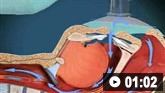
How to use bag-valve-mask apparatus to deliver ventilatory support to adults. Video demonstrates the two-person technique.
Enteral nutrition is generally a concern in the setting of encephalopathy, in which the patient is unable to obtain adequate nutrition due to an altered mental status. Therefore, enteral nutritional support with calorie-dense feeds should also be initiated early during the hospital course.
Blood glucose levels should be monitored every 1 to 2 hours by finger stick to assess for hypoglycemia. Hypoglycemia should be corrected with intravenous glucose infusion, with a glycemic target of 140 mg/dL.[51] Serum electrolytes, including sodium, phosphate, potassium, and magnesium, should be monitored at least twice daily and corrected aggressively.
Other routine laboratory studies such as coagulation activity, blood cell counts, and liver enzymes should also be monitored closely at regular intervals. Surveillance cultures from blood, urine, and sputum should be obtained periodically given the high risk of bacterial and fungal infection. The use of prophylactic antimicrobials has not been shown to affect clinical outcome.[4][8][103]
Proton-pump inhibitors or H2 antagonists are administered as prophylaxis for gastrointestinal bleeding.[8]
Lactulose and rifaximin are not used in the treatment of hepatic encephalopathy in ALF.[51]
Acetaminophen overdose or mild to moderate (grade 1 or 2) encephalopathy
Acetaminophen overdose is the most common cause of ALF in the US and Western Europe.[7] Determination of whether acetaminophen is responsible for ALF in an individual case is the most important factor to be addressed upon presentation.[104] Acetaminophen overdose is associated with depletion of hepatic glutathione stores and accumulation of a toxic intermediate, N-acetyl-p-benzoquinone imine, leading to direct hepatocyte injury. Restoration of glutathione synthesis within hepatocytes is dependent on cysteine, which may be administered in the form of acetylcysteine.[22]
In patients with acetaminophen overdose in whom ingestion is known to have occurred within 4 hours, a single-dose activated charcoal can be given.[4] Acetylcysteine therapy should be given in all cases of acetaminophen overdose regardless of the dose or timing of acetaminophen ingestion.[29] Acetylcysteine therapy should continue until endpoints such as improvement of hepatic function by clinical and laboratory parameters have been achieved.[22][99] See Acetaminophen overdose (Management approach).
One prospective placebo-controlled randomized clinical trial in patients with non-acetaminophen ALF reported a significant survival benefit in patients with grade 1 or 2 hepatic encephalopathy who received acetylcysteine versus placebo.[105] Consequently, acetylcysteine therapy is recommended for patients with ALF mild to moderate hepatic encephalopathy, even in the absence of acetaminophen ingestion.[104] Acetylcysteine may improve outcomes in non-acetaminophen ALF through mechanisms involving a reduction in expression of pro-inflammatory cytokines, such as IL-17, and decreased hepatocyte necrosis.[106][107] Based on available evidence, the American College of Gastroenterology recommends using intravenous acetylcysteine in patients with non-acetaminophen ALF.[4]
Other disease-specific therapies
These should be considered once the etiology of ALF has been established.[8][99] Some potential etiologies of ALF have specific therapies that may have an impact on clinical outcomes, including intravenous acyclovir for herpes simplex hepatitis and expedient delivery of the fetus in acute fatty liver of pregnancy or the hemolysis, elevated liver enzymes, and low platelet (HELLP) syndrome.
In cases of suspected Amanita phalloides mushroom poisoning, gastric lavage, activated charcoal, intravenous fluid resuscitation, intravenous penicillin-G, and acetylcysteine should be given.[4] Contact the poison control center for guidance. Silymarin (milk thistle) therapy has been described in Amanita phalloides poisoning, but no conclusive evidence supports its use.
Antiviral therapy for acute hepatitis B may also potentially have benefit and should be considered in ALF, although studies evaluating this are limited.[108]
[ ![]() ]
Entecavir or tenofovir are the preferred agents. No specific therapy is currently recommended for acute hepatitis A.
]
Entecavir or tenofovir are the preferred agents. No specific therapy is currently recommended for acute hepatitis A.
Patients presenting with acute Budd-Chiari syndrome should be considered for prompt initiation of anticoagulation therapy. Hepatic vein angioplasty with stent or transjugular intrahepatic portosystemic shunt placement may be considered in patients not responding to anticoagulation.[4] However, some may ultimately require liver transplantation. One multicenter case series comprising patients with ALF caused by Budd-Chiari syndrome noted that most were associated with a hypercoagulable state and that early initiation of anticoagulation therapy may be associated with improved survival.[109]
In contrast, some causes of ALF are associated with relatively poor outcomes despite administration of specific therapies. Acute Wilson disease with ALF is associated with high mortality despite measures to decrease serum copper levels, including plasmapheresis, continuous veno-venous hemofiltration, albumin dialysis, or plasma exchange. Chelation therapy for Wilson disease in the setting of ALF is generally ineffective, may be associated with hypersensitivity, and is not recommended.
In acute presentations of autoimmune hepatitis resulting in ALF, corticosteroids may have some benefit, as data suggest that patients treated with corticosteroids may have a higher rate of spontaneous recovery.[7] However, data have also demonstrated no significant survival benefit associated with corticosteroid use and suggest that its use in patients with ALF and high Model for End-Stage Liver Disease (MELD) scores could potentially lead to an increased risk of mortality.[110] In the absence of clear prospective data, the potential benefit of corticosteroids in the setting of ALF is uncertain.
Liver transplantation
Liver transplantation should be considered in all patients with ALF.[64] Prognostic scoring systems are helpful to identify patients at high risk of mortality, but have limitations and should not be relied upon to select patients for transplantation.[8] Therefore, measures should be taken early during the hospital course to prepare a patient with ALF for transfer to a nearby liver transplant center. Liver transplantation has a major impact on patient survival in ALF. According to reports, patients with ALF who undergo liver transplantation have an overall 5-year survival rate of 93%.[111]
Wilson disease should be suspected in any patient presenting with ALF with nonimmune hemolytic anemia including acute intravascular hemolysis. These patients should be urgently evaluated for liver transplantation.[42]
Patients with ALF who fulfill listing criteria according to the United Network for Organ Sharing (UNOS) may be assigned category Status 1A and listed with top priority for liver allocation. Criteria for UNOS Status 1A designation include:
Age >18 years, life expectancy without a liver transplant of <7 days, onset of encephalopathy within 8 weeks of the first symptoms of liver disease, absence of preexisting liver disease, admission to an intensive care unit, and 1 of the following:
Ventilator dependence, requirement of renal replacement therapy, or INR >2.0.
Patients with acute fulminant Wilson disease may also be given Status 1A priority.
Contraindications to liver transplantation for ALF include severe cardiac or pulmonary disease, AIDS, extrahepatic malignancy, metastatic hepatocellular carcinoma, intrahepatic cholangiocarcinoma, uncontrolled sepsis, irreversible neurologic complications (e.g., brain death, intracerebral hemorrhage, intractable sustained raised intracranial pressure), ongoing alcohol or illicit substance misuse, and lack of an adequate social support system.[48]
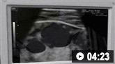
Ultrasound-guided insertion of a non-tunnelled central venous catheter (CVC) into the right internal jugular vein using the Seldinger insertion technique.
Use of this content is subject to our disclaimer