Aetiology
The aetiology of IAA varies according to the source of the infection and the status of the patient's immune system.
IAA that develops secondary to a localised peritonitis is usually due to a perforated viscus or a direct inoculation after trauma or recent surgery. IAA are commonly secondary to appendicitis (59%), diverticulitis (26%), and surgical procedures (11%).[1] Abscess in solid organs may be secondary to haematogenous seeding, whether through the portal system in the case of hepatic abscess or from various extra-abdominal locations when bacteraemia occurs. Infections associated with intra-peritoneal sepsis are polymicrobial in half of patients, and caused by a single isolate 25.7% of the time.[8] Although most cases of IAA are thought to be secondary to infection, microbiological confirmation of IAA is inconsistent, and bacterial growth is absent in about 26% of cases.[1]
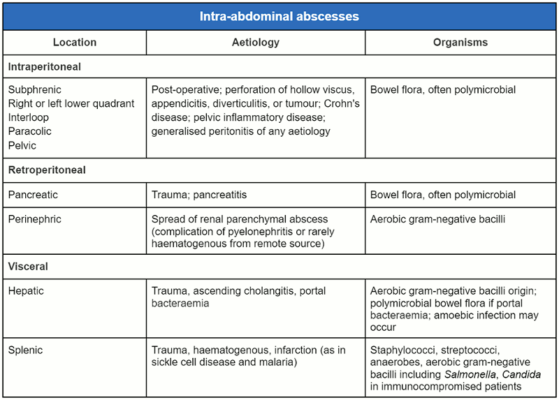
IAA are also classified as intra-peritoneal, retro-peritoneal, or visceral. Intraperitoneal (subphrenic, right or left lower quadrant, interloop, paracolic, pelvic) IAA are caused by bowel flora and are often polymicrobial.[9] They can occur post-operatively or as a result of perforation of a hollow viscus, appendicitis, diverticulitis, tumour, Crohn's disease, pelvic inflammatory disease, or generalised peritonitis of any aetiology. Retroperitoneal IAA can be either pancreatic, as a result of trauma or pancreatitis, or perinephric secondary to the spread of a renal parenchymal abscess, a complication of pyelonephritis or, rarely, due to haematogenous spread from a remote source. Pancreatic and perinephric abscesses are usually caused by bowel flora (often polymicrobial) and aerobic gram-negative bacilli respectively.[10][11]
Visceral IAA involve the liver or spleen. Splenic abscesses occur as a result of trauma, haematogenous spread, or infarction secondary to sickle cell disease or malaria. They are caused by staphylococci, streptococci, anaerobes, aerobic gram-negative bacilli (e.g., Salmonella), and Candida in immunosuppressed patients.[12][Figure caption and citation for the preceding image starts]: Classification of intra-abdominal abscesses (intraperitoneal, retroperitoneal, or visceral)From the collection of Dr Ali F. Mallat and Dr Lena M. Napolitano; used with permission [Citation ends].
Pathophysiology
IAA formation follows the same course as that of any other abscess. In a localised peritonitis, the host defence mechanism isolates the inflammatory reaction, but the debris and the oedema form a collection that grows progressively and becomes walled off and isolated.[9] Due to the non-vascularised structure, as well as the significantly acidotic medium, antibiotics are ineffective.[13] Active mechanical drainage of the abscess is the necessary treatment when the abscess increases in size.
Many IAA are polymicrobial, and aerobic and anaerobic gastrointestinal organisms such as Escherichia coli and Bacteroides often predominate.[14] The abscess environment often presents special challenges for antimicrobial therapy.[14] Abscesses have a low oxidation-reduction potential and low pH as a consequence of limited vascularity and poor perfusion, anaerobic conditions, and dying tissue. High bacterial concentrations tend to depress oxygen-dependent phagocytosis and killing of bacteria by neutrophils, and to suffuse the confined space with high concentrations of beta-lactamase enzymes. Antibiotic penetration into the abscess is limited not only by poor perfusion but also by mechanical barriers such as fibrin clots and the abscess wall.[14][15] Abscesses can be drained by percutaneous, laparoscopic, or open surgical techniques. The decision for the optimal approach for drainage often depends on the accessibility of the abscess percutaneously, as well as the degree of patient systemic illness.
Classification
Clinical anatomical classification
IAA can be classified based on the location of the abscess (such as interloop, sub-diaphragmatic, paracolic, or pelvic abscess) or in relation to the involved abdominal organ (such as hepatic, splenic, pancreatic, appendiceal, or diverticular abscess).
Assessing risk of adverse outcome and treatment failure[2]
Assess phenotypic and physiological factors:
Signs of sepsis
Extremes of age
Comorbidities
Extent of abdominal infection and adequacy of source control
Presence of resistant or opportunistic pathogen.
Characterise patients as either low or high risk for treatment failure or mortality.
Assess for community-acquired or health care-acquired infection.
Patients with Surviving Sepsis Campaign criteria for sepsis or septic shock, and those with APACHE II score greater than or equal to 10, are at higher risk.
Prolonged length of hospitalisation prior to surgery for intra-abdominal infection.
Patients with diffuse peritonitis.
Patients with delayed source control.
Use of this content is subject to our disclaimer