Images and videos
Images


Assessment of aphasia
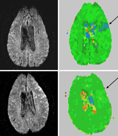
Diffusion-weighted image (top left) and perfusion-weighted image (top right) at day 1 of stroke; arrow points to area of hypoperfusion (blue). Lower panel shows corresponding views at day 2
From the collection of Dr Argye E. Hillis; used with permission
See this image in context in the following section/s:
Assessment of aphasia
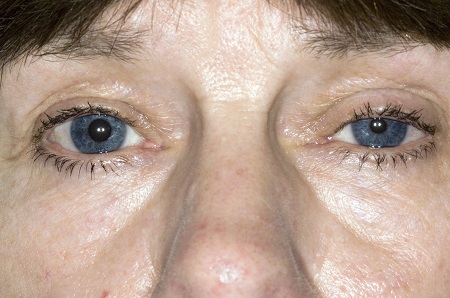
Ptosis and miosis affecting the patient's left eye.
Reproduced with permission from science photo library.
See this image in context in the following section/s:

Assessment of aphasia
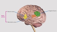
Broca’s area, Wernicke’s area and the angular gyrus.
Created by the BMJ Knowledge Centre
See this image in context in the following section/s:

Assessment of aphasia
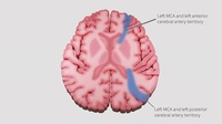
Watershed areas between the anterior, middle and posterior cerebral artery territories.
Created by the BMJ Knowledge Centre.
See this image in context in the following section/s:
Videos
 Diagnostic lumbar puncture in adults: animated demonstration
How to perform a diagnostic lumbar puncture in adults. Includes a discussion of patient positioning, choice of needle, and measurement of opening and closing pressure.
Use of this content is subject to our disclaimer