Approach
Historic time course aids in the differential diagnosis of aphasia. Sudden-onset or rapid progression implies stroke or other vascular cause. A slowly progressive course leads to consideration of degenerative, neoplastic, or neuroimmunological causes.
History
A sudden onset of aphasia in an otherwise healthy adult is most likely to be vascular (ischaemic stroke or intracerebral haemorrhage). However, tumour, infection, or other lesion limited to a single vascular territory will often mimic the stroke syndrome.
Considerations of pace of onset and coincident disease may broaden the differential diagnosis as to the cause.[38]
Rapid onset with poor nutrition will lead to suspicion of Wernicke's encephalopathy where it mimics aphasia.
Rapid-onset fluent aphasia with severe impairments in word meaning should be treated as herpes encephalitis until proven otherwise.[23]
Aphasia due to seizure or migraine may appear in patients with a history of epilepsy or migraine headaches.
Aphasia is an atypical presenting feature in prion disease (e.g., Creutzfeldt-Jakob disease). Isolated aphasia may be of short or prolonged duration.[32][33][34]
Weight loss can be a sign of cancer or nutritional deficiency, raising the suspicion for tumour or Wernicke's encephalopathy.
Impairment of language over at least 2 years followed by the onset of other cognitive or behavioural deficits suggests a neurodegenerative disorder, such as Alzheimer's disease or primary progressive aphasia.[26][27]
General examination
Non-specific manifestations may point toward the aetiology of aphasia.
Fever or tachycardia may signal an infectious cause such as herpes encephalitis or brain abscess.
Neck stiffness occurs with meningoencephalitis or subarachnoid haemorrhage.
Depressed, apathetic, or manic affect is common after stroke or in dementia.
Cardiac arrhythmia, carotid bruit, and peripheral pulse deficit suggest ischaemic stroke, central nervous system Lyme disease, or carotid stenosis.
Traumatic aphasia may be accompanied by typical findings of head injury such as bruises, fractures, bleeding, or watery nasal discharge (cerebrospinal fluid [CSF] rhinorrhoea).
Ophthalmic signs such as ptosis, miosis, visual field cuts, or ophthalmoplegia may also occur.
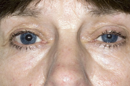
Unilateral ptosis and miosis are signs of carotid dissection as a cause of stroke.[Figure caption and citation for the preceding image starts]: Ptosis and miosis affecting the patient's left eye.Reproduced with permission from science photo library. [Citation ends].
Ophthalmoplegia is associated with Wernicke's encephalopathy and tauopathies (neurodegeneration resulting from tau protein neurofibrillary tangles) that can cause progressive non-fluent aphasia.
An intranuclear ophthalmoplegia suggests multiple sclerosis.
Visual field deficits are common in stroke and migraine.
Neurological examination
Neurological deficits may indicate the relative extent of damage to the brain. Stance and gait may be affected by truncal rigidity or hemiparesis of the leg. Sensory deficits on the right often accompany aphasia due to left parietal or thalamic lesions of any cause.
Several aspects of language are tested to distinguish between various aphasia syndromes, associated with particular vascular territories.[39] Keep in mind that any lesion within a particular vascular territory (not just stroke) can cause symptoms of the associated aphasia syndrome. Impairments of the following language domains provide clues to the location of damage:
Quality of spontaneous speech (e.g., agrammatic speech output in Broca's aphasia)
Naming (deficits typical of all aphasias, but also of dementia)
Repetition (disproportionately impaired in conduction aphasia and disproportionately spared in transcortical aphasias)
Comprehension of semantically reversible versus non-reversible sentences (both impaired in Wernicke's aphasia; only the former impaired in Broca's aphasia)
Reading and writing (deficits associated with all aphasias, but can also occur in isolation).
Evaluation of cognition will help distinguish aphasia from more diffuse disorders such as dementia, in which there is derangement of:
Attention span
Visuospatial skills
Recent and remote memory
Executive functions
Social behaviour or comportment.
Upper motor neuron and cranial nerve findings may indicate a vascular or non-vascular aphasia:
Upper motor neuron facial weakness often accompanies stroke.
Multiple sclerosis and the tauopathies that cause progressive non-fluent aphasia can cause central (upper motor neuron) cranial nerve dysfunction.
Other cranial nerve deficits should raise suspicion for a systemic aetiology of aphasia (e.g., infection or tumour).
Lower motor neuron deficits are generally features of aphasia of non-vascular aetiology:
Lyme disease
Carcinomatous or lymphomatous meningitis
Sarcoidosis
Aphasia dysarthria motor neuron disease (amyotrophic lateral sclerosis/frontotemporal dementia).
Ataxia is associated with true aphasia in the case of:
Multiple strokes or multiple lesions in multiple sclerosis
Sarcoidosis
Neoplastic disease
Trauma
Wernicke's encephalopathy
Some rare degenerative diseases.
Imaging
Non-contrast computed tomography (CT) of the head
Brain imaging should be completed rapidly to distinguish between ischaemic stroke, intracerebral haemorrhage, and subdural haematoma.[21][22][37][41]
Magnetic resonance imaging (MRI) with diffusion weighted imaging
Angiography
Detailed vascular images with CT angiography or MR angiography are adjuncts to initial studies in cases where a vascular cause is suspected.[40]
Conventional invasive brain angiography (catheter intra-arterial digital subtraction angiography [DSA]) also has a role; it is recommended in patients with spontaneous ICH and a CT or MR angiogram suggestive of a macrovascular cause (e.g., arterio-venous malformation or aneurysm).[37]
MRI with and without contrast when vascular aetiology is not suspected
Essential for a broader differential diagnosis, including demyelinating disease, cancer, and infections.
CT or MRI to evaluate for asymmetric left hemisphere atrophy
Can help distinguish types of primary progressive aphasia from other degenerative diseases.
Positron emission tomography (PET) or single photon emission computed tomography
Focal hypoperfusion or hypometabolism can also be helpful in distinguishing subtypes of primary progressive aphasia from each other and from other degenerative conditions.[42]
Multiple amyloid PET tracers (e.g., florbetapir, florbetaben, flutametamol) specifically bind to fibrillar amyloid plaques; an elevated signal is supportive of a diagnosis of Alzheimer’s disease.[43]
Specialised cardiac and vascular studies may be helpful in evaluation of aphasia
Echocardiogram may reveal vegetation or other source of cardioembolism and can direct therapy (i.e., anticoagulation).
Bubble study (saline contrast enhances visualisation of cardiac septal defects) may be helpful in detection of adult patent foramen ovale.[44]
Carotid Doppler may reveal common or internal carotid stenosis contributing to vascular insufficiency and stroke.[45]
Magnetic resonance angiography of neck/circle of Willis may identify occlusion contributing to vascular insufficiency and can direct specific therapy to the area of concern.
Chest x-ray and CT chest
May be diagnostic of hilar lymphadenopathy raising suspicion of sarcoidosis as aetiology of aphasia.
Ancillary studies
Lumbar puncture
Essential if an infectious cause is suspected on the basis of fever, tachycardia, nausea, or leukocytosis.
CSF can also be useful in diagnosing neoplastic causes, multiple sclerosis, and other neuroimmunological causes.
CSF evidence of amyloidosis may provide additional evidence favouring diagnosis of Alzheimer's disease.
 Diagnostic lumbar puncture in adults: animated demonstration
How to perform a diagnostic lumbar puncture in adults. Includes a discussion of patient positioning, choice of needle, and measurement of opening and closing pressure.
Electroencephalography
Critical if seizure is the suspected cause.
Can also be helpful in diagnosing herpes encephalitis and Creutzfeldt-Jakob disease.
Neuropsychological testing
Sometimes necessary to differentiate aphasia from dementia.
Electromyography
May reveal lower motor neuron lesions associated with amyotrophic lateral sclerosis.
Laboratory tests
Serum thiamine is requested if Wernicke's encephalopathy is suspected (e.g., in people with alcohol use disorder) and clinical signs are present, such as confabulation, diplopia, nystagmus, and ataxia. Presumptive treatment with intravenous thiamine should not be delayed pending the results of laboratory investigations.
Serum glucose should be checked in all patients with suspected stroke.
Full blood count should be requested when an infectious aetiology is possible.
Use of this content is subject to our disclaimer