Images and videos
Images


Assessment of vision loss
Central retinal vein occlusion: extensive retinal haemorrhages and dilated vessels
From Dr Prem S. Subramanian's personal collection; used with permission
See this image in context in the following section/s:

Assessment of vision loss
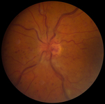
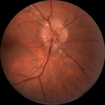
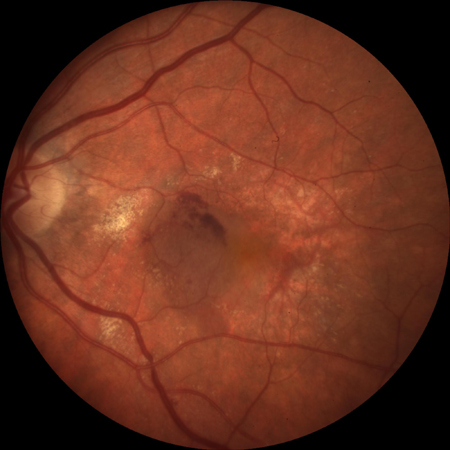
Right optic nerve with disc swelling
From Dr Prem S. Subramanian's personal collection; used with permission
See this image in context in the following section/s:
Assessment of vision loss
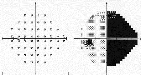
Left eye of patient with homonymous field defect
From Dr Prem S. Subramanian's personal collection; used with permission
See this image in context in the following section/s:

Assessment of vision loss
Fluorescein angiogram showing classic choroidal neovascularisation with early hyperfluorescence
From Dr Prem S. Subramanian's personal collection; used with permission
See this image in context in the following section/s:

Assessment of vision loss
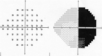
Right eye of patient with homonymous field defect
From Dr Prem S. Subramanian's personal collection; used with permission
See this image in context in the following section/s:
Assessment of vision loss
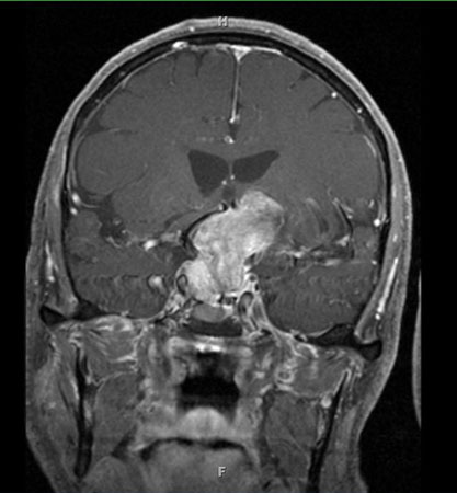
Pituitary apoplexy: large suprasellar mass with heterogeneous gadolinium enhancement (T1-weighted MRI)
From Dr Prem S. Subramanian's personal collection; used with permission
See this image in context in the following section/s:

Assessment of vision loss
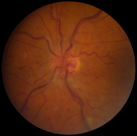
Segmental optic disc swelling and haemorrhage seen in non-arteritic ischaemic optic neuropathy
From Dr Prem S. Subramanian's personal collection; used with permission
See this image in context in the following section/s:
Assessment of vision loss
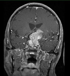
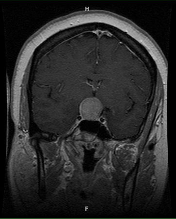
Pituitary tumour: homogenous suprasellar mass elevating and compressing optic chiasm (MRI)
From Dr Prem S. Subramanian's personal collection; used with permission
See this image in context in the following section/s:
Assessment of vision loss
Sub-retinal haemorrhage and retinal elevation from sub-foveal neovascularisation
From Dr Prem S. Subramanian's personal collection; used with permission
See this image in context in the following section/s:

Assessment of vision loss
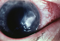
Corneal ulcer with epithelial defect and stromal infiltrate
From Dr Prem S. Subramanian's personal collection; used with permission
See this image in context in the following section/s:
Assessment of vision loss
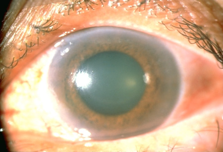
Angle-closure glaucoma: the pupil is mid-dilated, and the cornea is oedematous and cloudy, as indicated by the dulled and irregular light reflex
From Dr Prem S. Subramanian's personal collection, used with permission
See this image in context in the following section/s:

Assessment of vision loss
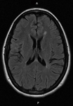
Typical white matter lesions indicative of multiple sclerosis risk (MRI)
From Dr Prem S. Subramanian's personal collection; used with permission
See this image in context in the following section/s:
Use of this content is subject to our disclaimer