Images and videos
Images


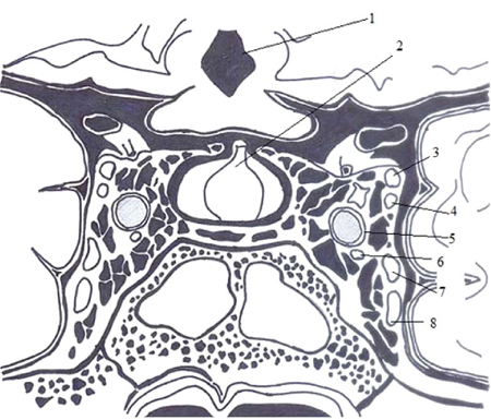
Cavernous sinus thrombosis
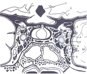
Anatomy of the cavernous sinus: (1) third ventricle, (2) pituitary gland, (3) oculomotor nerve, (4) trochlear nerve, (5) internal carotid artery, (6) abducens nerve, (7) ophthalmic branch of the trigeminal nerve, and (8) maxillary branch of the trigeminal nerve
Visvanathan V, et al. Reminder of important clinical lesson: ocular manifestations of cavernous sinus thrombosis. BMJ Case Rep. 2010; doi:10.1136. Used with permission
See this image in context in the following section/s:
Cavernous sinus thrombosis
Ocular manifestations of cavernous sinus thrombosis and their underlying pathology
Visvanathan V, et al. Reminder of important clinical lesson: ocular manifestations of cavernous sinus thrombosis. BMJ Case Rep. 2010; doi:10.1136. Used with permission
See this image in context in the following section/s:
Cavernous sinus thrombosis
Treatment of septic cavernous sinus thrombosis (CST)
From the collection of Dr Jayant Pinto, University of Chicago
See this image in context in the following section/s:
Cavernous sinus thrombosis
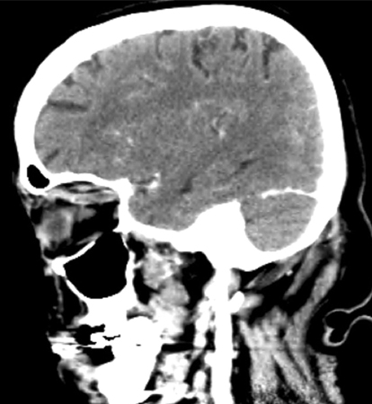
Sagittal CT scan of the head demonstrating an enlarged, tubular right superior ophthalmic vein
Jones RG, Arnold B. Sudden onset proptosis secondary to cavernous sinus thrombosis from underlying mandibular dental infection. BMJ Case Rep. 2009;2009. pii: bcr03.2009.1671. Used with permission
See this image in context in the following section/s:

Cavernous sinus thrombosis
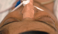
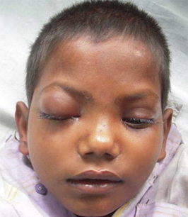
Proptosis of the right eye in a patient with cavernous sinus thrombosis secondary to dental infection
Jones RG, Arnold B. Sudden onset proptosis secondary to cavernous sinus thrombosis from underlying mandibular dental infection. BMJ Case Rep. 2009;2009. pii: bcr03.2009.1671. Used with permission
See this image in context in the following section/s:

Cavernous sinus thrombosis
Treatment of aseptic cavernous sinus thrombosis (CST)
From the collection of Dr Jayant Pinto, University of Chicago
See this image in context in the following section/s:
Cavernous sinus thrombosis
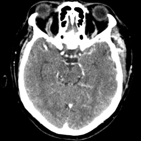
Post-contrast venous phase CT scan of the head (axial view) showing an enlarged 'S'-shaped right superior ophthalmic vein with associated proptosis
Jones RG, Arnold B. Sudden onset proptosis secondary to cavernous sinus thrombosis from underlying mandibular dental infection. BMJ Case Rep. 2009;2009. pii: bcr03.2009.1671. Used with permission
See this image in context in the following section/s:
Cavernous sinus thrombosis
Patient with bilateral cavernous sinus thrombosis. Note the bilateral proptosis, which is more marked in the right eye
Vidhate MR, et al. Bilateral cavernous sinus syndrome and bilateral cerebral infarcts: a rare combination after wasp sting. J Neurol Sci. 2011;301:104-106. Used with permission
See this image in context in the following section/s:
Use of this content is subject to our disclaimer