Approach
Presentation is usually in childhood, often in the first year of life. Thrombocytopenia is universal, but only one third of patients have the other features of immunodeficiency and eczema at presentation.[1] Attenuated WAS may initially be misdiagnosed as idiopathic thrombocytopenia. Diagnosis should be suspected if there is a positive family history of thrombocytopenia.
History and examination
Diagnosis is based on clinical suspicion. Children usually present with complications of thrombocytopenia, especially petechiae and easy bruising. Recurrent infections, particularly ear infections, occur, although this is less usual if the child presents in the first year of life. There may be a history of previous serious or opportunistic infection.[1] There is often a history of eczema. There may be a family history of bleeding or infant mortality. A wide variety of autoimmune syndromes occur later in the disease, which may affect 26% to 72% of patients at some point, and history of these should be sought.[17]
Examination findings usually include bruises and petechiae, and eczema of variable severity with petechiae or bleeding at sites of excoriation. Lymphadenopathy may be present, particularly if skin is inflamed. There may be evidence of past/current ear infections such as tympanic perforation. Failure to thrive may be present.[Figure caption and citation for the preceding image starts]: Eczema and bleeding in Wiskott-Aldrich syndromeFrom the collection of S. Burns and A. Thrasher; used with consent given by parents [Citation ends].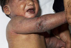
Initial diagnostic investigations
Initial investigation is a full blood count, and should include platelet volume. Thrombocytopenia with low platelet volume is universal unless splenectomy has been performed. WAS protein (WASp) analysis should be performed (either by Western blot, flow cytometry, or by fluorescence-activated cell sorter).[18] These are specialised tests only available at selected centres. WASp gene mutation analysis should also be performed as the definitive test for diagnosis.
Subsequent investigations
A full immune work-up is indicated, including immunoglobulin levels (IgG, IgM, and IgA), vaccine antibody responses (such as Hib, tetanus, and pneumococcal antibody levels), isohaemagglutinins, lymphocyte subset analysis, and T-cell stimulation assays (Pha and anti-CD3 stimulation).
Prior to commencing treatment with immunoglobulin, liver function tests should be performed and immunoglobulin levels (IgG, IgM, and IgA) repeated. Polymerase chain reaction (PCR) analysis for hepatitis C is also recommended as a baseline to document lack of infection prior to immunoglobulin (blood product) exposure. Serum and plasma should also be stored in case they are needed at a later date for specific pre-immunoglobulin antibody levels and PCR tests (e.g., if the patient develops immunoglobulin-transmitted infection). In general, adenovirus, Epstein-Barr virus, and cytomegalovirus PCR tests should be performed to document past or present infection; this is particularly important for patients intended for bone marrow transplant, when infection status is key for treatment planning. In the event of significant eczema, an assessment by a dermatologist may be required to determine triggering factors such as food allergy.
Use of this content is subject to our disclaimer