This page compiles our content related to dermatitis. For further information on diagnosis and treatment, follow the links below to our full BMJ Best Practice topics on the relevant conditions and symptoms.
Introduction
Relevant conditions
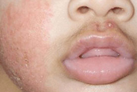
EczemaSigns & symptomsInvestigationsDifferentialsTreatment algorithm | go to our full topic on Eczema Commonly presents with dry, itchy skin. Typically there is erythema, scaling, vesicles, or lichenification in skin flexures. In infants, the extensor surfaces, cheeks, and forehead are preferentially affected.[1] Patients often have a personal or family history of other atopic diseases such as asthma or allergic rhinitis.[1][2][3] Eczema is a chronic, relapsing disease, and educating patients and their families is necessary so that they develop an understanding of basic skin care and how to avoid trigger factors.[4][5][6][7][Figure caption and citation for the preceding image starts]: Acute atopic dermatitis on the face of an infantPersonal collection of Dr A. Hebert [Citation ends]. |
|---|---|
Contact dermatitisSigns & symptomsInvestigationsDifferentialsTreatment algorithm | go to our full topic on Contact dermatitis Irritant contact dermatitis is caused by direct toxicity and can occur in any person without prior sensitisation. Allergic contact dermatitis is a delayed hypersensitivity reaction, which requires prior sensitisation.[8] Patients generally report pruritus, burning, erythema, swelling, and blistering with acute contact dermatitis, and pruritus, burning, erythema or hyperpigmentation, fissuring, and scaling with chronic contact dermatitis. |
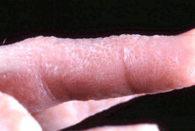
Dyshidrotic dermatitisSigns & symptomsInvestigationsDifferentialsTreatment algorithm | go to our full topic on Dyshidrotic dermatitis Recurrent crops of 1- to 2-mm vesicles, with pruritus on the palms, soles, and/or lateral aspects of the fingers. Pompholyx is a term often used synonymously with dyshidrotic dermatitis, but it is better used to describe more acute, severe eruptions of large bullae on the hands and feet.[9] The common exacerbating factor is irritation, as seen in frequent hand washing, hyperhidrosis, and stress. However, the underlying aetiology is unknown. [Figure caption and citation for the preceding image starts]: Dyshidrotic eczemaPhotograph courtesy of Dr Spencer Holmes [Citation ends]. |
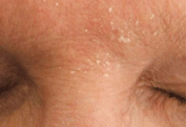
Seborrhoeic dermatitisSigns & symptomsInvestigationsDifferentialsTreatment algorithm | go to our full topic on Seborrhoeic dermatitis A common inflammatory skin disorder that usually manifests as erythema and scaling of the scalp, nasolabial folds, and occasionally central face and anterior chest. It tends to worsen with stress.[10] The adult scalp form is commonly termed dandruff or pityriasis capitis. Variable course that seldom completely subsides. An infant form (cradle cap) usually resolves within the first few months of life. [Figure caption and citation for the preceding image starts]: Seborrhoeic dermatitis, glabella, with scaling and mild erythemaPersonal collection of Dr Robert A. Schwartz [Citation ends]. |
Nappy rashSigns & symptomsInvestigationsDifferentialsTreatment algorithm | go to our full topic on Nappy rash Primarily an irritant contact dermatitis, nappy rash is inflammation of the skin in the area of the body covered by a nappy. It is most common in the first 2 years of life, but can occur in any person who routinely wears nappies. Recalcitrant nappy rash may signal secondary infection or underlying systemic or dermatological disorders, and requires further evaluation. |
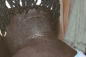
Lichen simplex chronicus (LSC)Signs & symptomsInvestigationsDifferentialsTreatment algorithm | go to our full topic on Lichen simplex chronicus (LSC) A common cutaneous disorder characterised by well-circumscribed erythematous, often hyperpigmented, patches and plaques of thickened lichenified skin. It most commonly occurs on the neck, ankles, scalp, pubis, vulva, scrotum, and extensor forearms as a result of chronic scratching and rubbing.[11]One or multiple LSC patches or plaques can arise on skin affected by an underlying dermatosis such as atopic dermatitis, allergic contact dermatitis, stasis dermatitis, superficial fungal (tinea and candidiasis) and dermatophyte infections, lichen sclerosis, viral warts, scabies, lice, arthropod bite, or cutaneous neoplasia.[11][12] LSC can be a difficult condition to treat, causing frustration in both the patient and physician.[11] [Figure caption and citation for the preceding image starts]: Secondary lichen simplex chronicus in the setting of atopic dermatitisPersonal collection of Dr Swick [Citation ends]. |
SunburnSigns & symptomsInvestigationsDifferentialsTreatment algorithm | go to our full topic on Sunburn An acute inflammatory reaction of the skin induced by over-exposure to ultraviolet (UV) radiation. Skin findings include erythema and oedema, with or without vesiculation, followed by desquamation. Symptoms include pain and/or pruritus. Acute sunburn is a self-limited condition and typically requires only supportive care. No current treatments can reverse UV-induced skin damage.[13] However, primary prevention is critical, as cellular damage caused by UV radiation is irreversible and may with time increase the risk of skin cancer. |
Assessment of pruritusDifferentials | go to our full topic on Assessment of pruritus The most subjective symptom in dermatology is itching, which may occur with or without visible skin lesions. A thorough history and complete physical examination are central to the evaluation of pruritus.[14] During clinical evaluation, it is important to identify a possible cause or disease responsible for itching, as well as determining the intensity and timeframe of the pruritus. |
Assessment of rash in childrenDifferentials | go to our full topic on Assessment of rash in children Rash in children is common. The differential diagnoses are extensive, ranging from self-limiting conditions (e.g., roseola) to life-threatening illnesses such as meningococcal disease. Initial considerations in evaluating a rash in children include its morphology, duration, and distribution. Age, sex, family history, medications, known allergies, and exposures are also of primary importance. |
Assessment of dermatological disorders in HIVDifferentials | go to our full topic on Assessment of dermatological disorders in HIV The dermatological manifestations of HIV are protean and often multiple in patients with HIV infection. HIV-specific dermatoses include HIV-related lipodystrophy, eosinophilic folliculitis, oral hairy leukoplakia, papular pruritic eruption of HIV, and HIV photodermatitis. Some skin diseases that appear in non-HIV-infected populations may have altered presentation in people with HIV. Seborrhoeic dermatitis occurs with strikingly increased prevalence in HIV infection.[15][16] Atopic dermatitis has a high prevalence in adult as well as paediatric populations with HIV.[17] |
Contributors
Authors
Editorial Team
BMJ Publishing Group
Disclosures
This overview has been compiled using the information in existing sub-topics.
Use of this content is subject to our disclaimer