Images and videos
Images
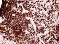


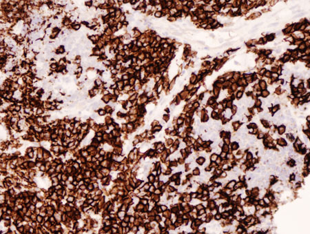
MALT lymphoma
Lung MALT lymphoma: parts of this tumour demonstrate striking plasmacytic differentiation (CD138 staining, ×200)
From the collections of Dr R. Joshi and Dr C. McNamara; used with permission
See this image in context in the following section/s:
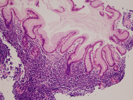
MALT lymphoma
Gastric MALT lymphoma: normal gastric epithelium distorted by a neoplastic infiltrate of lymphocytes extending into the superficial gastric epithelium (haematoxylin and eosin [H&E] staining, ×200)
From the collections of Dr R. Joshi and Dr C. McNamara; used with permission
See this image in context in the following section/s:
MALT lymphoma
Gastric MALT lymphoma: infiltration of the gastric epithelium by neoplastic B-lymphocytes (CD20 staining, ×200)
From the collections of Dr R. Joshi and Dr C. McNamara; used with permission
See this image in context in the following section/s:

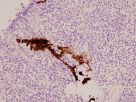
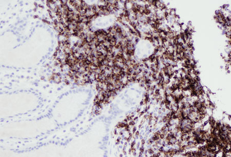
MALT lymphoma
Lung MALT lymphoma: residual respiratory epithelium has been distorted by infiltrating lymphocytes; the lymphoepithelial lesion (cytokeratin staining, ×200)
From the collections of Dr R. Joshi and Dr C. McNamara; used with permission
See this image in context in the following section/s:
MALT lymphoma
Lung MALT lymphoma: lung parenchyma has been replaced by a neoplastic infiltrate of small lymphocytes; a follicle surrounded by neoplastic marginal zone cells can be recognised in the centre of the image (haematoxylin and eosin [H&E] staining, ×200)
From the collections of Dr R. Joshi and Dr C. McNamara; used with permission
See this image in context in the following section/s:

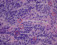
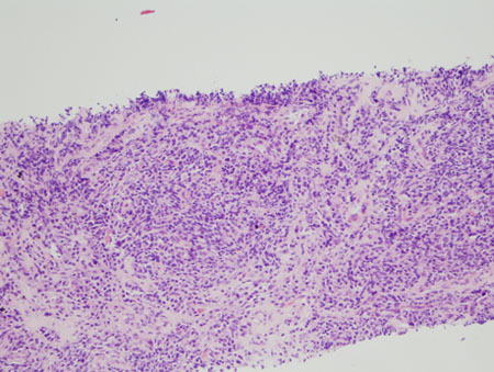
MALT lymphoma
MALT lymphoma has a polymorphous lymphoid infiltrate that can include small lymphocytes, monocytoid B cells, centrocyte-like cells and cells with plasmacytoid differentiation. Scattered immunoblasts and centroblast-like cells may also be seen
Hollie N, et al. J Clin Pathol. 2020; 73: 378-83; used with permission
See this image in context in the following section/s:

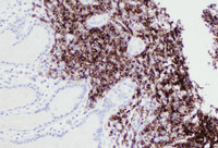
MALT lymphoma
Infiltrate of lymphoid cells in the lung, confirming their B-cell origin (CD20 staining, ×200)
From the collections of Dr R. Joshi and Dr C. McNamara; used with permission
See this image in context in the following section/s:
Use of this content is subject to our disclaimer