Approach
A diagnostic evaluation differs greatly depending on whether the urticaria is acute or chronic, and whether a patient has angio-oedema in the absence of urticaria.
Diagnosis of acute urticaria (i.e., episodes lasting <6 weeks) is predominantly based on history and physical examination, while a diagnosis of chronic urticaria (i.e., episodes lasting ≥6 weeks) may involve additional laboratory testing to identify treatable associated conditions or to exclude other unusual conditions such as urticarial vasculitis.
When there is angio-oedema in the setting of urticaria (occurs in up to 40% of patients), the general approach to diagnosis is like that for urticaria alone.
Angio-oedema occurring in the absence of urticaria requires a more directed evaluation aimed at identifying cases of hereditary angio-oedema, acquired angio-oedema, or drug-induced angio-oedema.
It is essential that any patient presenting with angio-oedema affecting the neck, face, tongue, or lips have their airway assessed and measures taken to ensure that it is protected before any further evaluation or diagnostic work-up is performed. If available, flexible fibre-optic laryngoscopy can rapidly determine the extent of involvement of the base of the tongue or the larynx. This can determine the most appropriate airway management strategy.[26][Figure caption and citation for the preceding image starts]: Angio-oedema of the lips in a patient who also has urticariaFrom the collection of Stephen Dreskin, MD, PhD [Citation ends].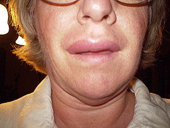
Acute urticaria: history
History-taking should aim to elicit the nature of the lesions and any specific triggers.
Patients are often able to identify triggers such as foods or drugs. Questions should explore potential relationships between the urticarial lesions and changes or additions to drugs (including non-prescription, herbal, and topical agents), ingestion of food products, contact exposures, physical triggers, insect bites/stings, or any other suspected agents.
Questioning should cover the size, shape, frequency, and duration of the lesions, as well as subjective symptoms of pruritus or pain. Any association with angio-oedema and response to any previous therapy should also be explored.[Figure caption and citation for the preceding image starts]: Typical lesions seen in acute or chronic urticariaFrom the collection of Stephen Dreskin, MD, PhD [Citation ends].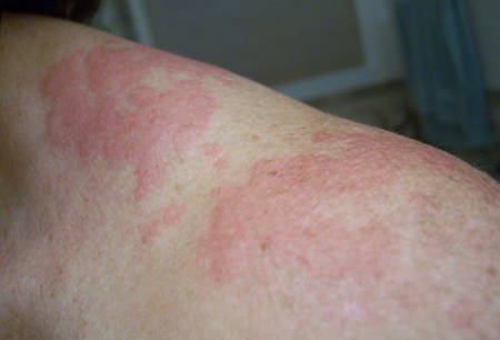
Up to 90% of patients with anaphylaxis have symptoms of urticaria and angio-oedema.[27] Anaphylaxis is a life-threatening multi-system disorder. It should be excluded promptly by enquiring about other symptoms of anaphylaxis including: respiratory symptoms (dyspnoea, wheeze, rhinitis); gastrointestinal symptoms (e.g., nausea, vomiting, abdominal pain, diarrhoea); diaphoresis, dizziness, retrosternal pain, and headache.[27][28]
Acute urticaria: physical examination
Physical examination should be thorough and complete, and directed at establishing underlying causes of the urticaria. Examination of the skin should include testing for dermatographism by stroking or scratching the skin. Tests for inducible urticaria should also be considered if indicated by the history.
Lesions can be characterised as typical or atypical. Typical lesions are oedematous pink or red wheals of variable size and shape, with surrounding erythema. Multiple pinpoint lesions are characteristic of cholinergic urticaria. Atypical lesions are purpuric, non-blanchable, and palpable. They may also leave residual pigmented skin. Atypical lesions are associated with urticarial vasculitis.
Signs of shock, wheeze, stridor, and respiratory distress should raise suspicion of anaphylaxis and prompt immediate administration of intramuscular adrenaline (epinephrine).
Acute urticaria: diagnostics
If the history suggests potential triggers of the urticaria, these need to be formally assessed. Triggers frequently include food items or drugs. It is important not to overlook herbal medicines or dietary supplements. Any drug that could be a trigger is replaced with a chemically unrelated alternative. If urticaria persists despite elimination of the suspected inciting agent, this is unlikely to be the cause.
Testing for a broad panel of food or environmental allergens is generally ineffective and is not recommended. However, targeted testing for specific food or other allergens based on the history can provide greater diagnostic usefulness. Serological testing is generally preferred to skin prick testing, because the development of urticaria may confound interpretation of the skin test results.
Laboratory tests are not indicated in cases of acute urticaria, unless there are findings on history and physical examination that would indicate the lesions were atypical.[1] This should prompt evaluation for urticarial vasculitis. An elevated erythrocyte sedimentation rate or C-reactive protein may help distinguish urticarial vasculitis from urticaria. If there is high suspicion of urticarial vasculitis, a skin biopsy should be considered.
Chronic urticaria: history
As with acute urticaria, evaluation of chronic urticaria begins with a detailed history and physical examination. Because these patients frequently do not have lesions present at the time of the consultation, it is imperative to establish whether the lesions are truly urticaria. Asking the patient to photograph the lesions can be helpful in this regard.[29]
Key features include the size, shape, frequency, and duration of the lesions, as well as subjective symptoms of pruritus or pain.
Any association of the urticaria with angio-oedema, and the response to any previous therapies, should also be explored. While patients can often identify precipitating triggers for acute urticaria, it is very uncommon for patients to be able to identify triggers in chronic urticaria. In more than 90% of cases of chronic urticaria, neither the patient nor the physician is able to identify a trigger.[30] This can be a source of frustration, but this can be helped by giving the patient a thorough understanding of the pathophysiology of the disease.
Chronic urticaria: physical examination
Physical examination should be complete and directed at establishing potential causes of the urticaria and any other underlying illnesses. With chronic urticaria in particular, evaluation of potential inducible causes of the urticaria should form part of the examination:
Dermatographism is tested for by stroking or scratching the skin.
Cholinergic urticaria, which often occurs following exercise or exposure to heat, can present as diffuse erythema or as multiple, small lesions a few millimetres in diameter.
Cold urticaria is assessed by placing an ice cube on the skin for 5 minutes and observing for an urticarial lesion as the skin re-warms.
Delayed pressure urticaria can be tested for by placing a sand bag over the patient's shoulder for 15 to 30 minutes. The patient should then be instructed to observe for urticaria for 4 to 6 hours after the sand bag is removed.
Aquagenic urticaria can be tested for by applying a water compress to the skin or by immersing a limb under water. Exposure to water for up to 30 minutes may be needed to maximise sensitivity. In addition, although water of any temperature can provoke aquagenic urticaria, using a compress or bath at room temperature helps to minimise confusion with cold or local heat urticarias.
Other inducible urticarias, such as solar urticaria, can also be tested for by controlled exposure to the relevant physical stimuli.[31]
Test responses may be blunted if individuals are taking antihistamines concurrently.
Chronic urticaria: diagnostics
If no cause of the chronic urticaria can be established by the history and physical examination, it is then reasonable to consider a directed laboratory evaluation. This is particularly important to exclude urticarial vasculitis in patients who have urticarial lesions with atypical features (e.g., pain rather than pruritus, lesions lasting longer than 24 hours, residual bruising, non-blanching lesions).
The following tests are recommended in all patients with chronic spontaneous urticaria:[1]
FBC with differential
C-reactive protein (CRP) and/or erythrocyte sedimentation rate (ESR).
In most cases of chronic urticaria, an underlying cause cannot be identified from a careful history, and physical examination is normal. In these cases, further testing is not required.[32]
Additional diagnostic testing may be appropriate if a concomitant disorder associated with chronic urticaria is suspected and may include:[1][32][33]
Thyroid-stimulating hormone (TSH): only ordered if history or examination suggests thyroid dysfunction.
Antinuclear antibody (ANA): only ordered if history or examination suggest concomitant autoimmune disease (e.g., systemic lupus erythematosus [SLE], dermatomyositis, polymyositis, Sjogren syndrome, or Still's disease).[33]
Allergen avoidance diet: may be considered for 2 to 3 weeks if the history suggests food pseudoallergy (non-immunoglobulin E [IgE]-mediated hypersensitivity to food pseudoallergens). These include some artificial food additives, naturally occurring salicylic acid, and aromatic volatile compounds in herbs, wine, and tomatoes.[34]
Patients may strongly believe that certain foods are the cause of their urticaria, and this belief can sometimes cause difficulty for the clinician. In this situation, the best approach is to ask the patient to maintain a detailed food diary. If any foods are found to correlate with episodes of urticaria, they can be eliminated from the diet and the patient observed for resolution of the urticaria. Foods can then be gradually reintroduced into the diet as tolerated. If angio-oedema has been a feature of the patient's condition, this reintroduction should be performed in a setting equipped to manage allergic reactions, such as a doctor's surgery. However, it should be stressed that foods typically do not cause chronic urticaria.
Serum tryptase: ordered if history or clinical examination suggests systemic mastocytosis.
Skin biopsy: usually only performed if the condition does not respond to antihistamines or there is concern for urticarial vasculitis.
Skin testing for immediate hypersensitivity: should only be considered if IgE-mediated food allergy is suspected as a cause. One study found that nearly 40% of people with chronic urticaria had a positive skin prick test, but in double-blind, placebo-controlled provocation tests, administration of the allergen did not provoke urticaria symptoms in any patients.[9] It should be stressed that foods typically do not cause chronic urticaria and performing skin testing for a broad panel of food allergens is not recommended.
Routine or intensive screening programmes for causes of urticaria are not recommended.[1][35]
Several chronic infections have been reported as causes of chronic urticaria: for example, hepatitis B and C, Epstein-Barr virus, and Helicobacter pylori. Evidence does not support testing for these conditions in a patient who has an otherwise unremarkable history and physical examination. Evidence is lacking that treatment of the infection, based on abnormal test results, relieves symptoms or alters the clinical course of chronic urticaria.[33]
Angio-oedema without urticaria
Isolated angio-oedema in the absence of associated urticaria requires an alternative diagnostic approach. A thorough history and physical examination still forms the basis of diagnosis, but particular attention is paid to the drug history and family history with the aim of identifying cases of drug-induced angio-oedema, hereditary angio-oedema, or acquired angio-oedema.
Examination should include vital signs, followed by rapid assessment of the head and neck, skin, and abdomen. Stridor, voice hoarseness, and the presence of angio-oedema in the lips, tongue, soft palate, or posterior pharynx should be noted. If available, flexible fibre-optic laryngoscopy can rapidly determine the extent of involvement of the base of the tongue or the larynx. This can determine the most appropriate airway management strategy.[26]
Skin examination will reveal the extent and distribution of the angio-oedema. Abdominal pain can occur in histaminergic and non-histaminergic angio-oedema. Bowel wall oedema in patients with hereditary angio-oedema may cause severe abdominal pain, guarding, and rebound tenderness, mimicking an acute surgical abdomen.[26]
Laboratory tests that apply to cases of angio-oedema without urticaria are based on complement level and function: C4 level, C1 esterase inhibitor level, C1 esterase inhibitor function, and C1q level. Initial screening should include C4 level only, unless there is a compelling family history for hereditary angio-oedema. C4 level cannot be relied upon to confirm or exclude a diagnosis of hereditary angio-oedema.[36]
Drug-induced angio-oedema
Swelling can involve any body part, but typically involves the hands, feet, and face. Tongue swelling and airway closure are also commonly reported.
ACE inhibitors are a common cause, with angio-oedema occurring in up to 0.7% of individuals on these drugs.[11] Although ACE inhibitor-induced angio-oedema typically occurs within days of initiating ACE inhibitor therapy, there are some individuals who only experience it after several years of treatment.
Non-steroidal anti-inflammatory drugs (NSAIDs) and antibiotics are also common causes of drug-induced angio-oedema. Any drug considered a potential cause should be discontinued and/or replaced with a chemically unrelated agent and the patient observed for resolution of the angio-oedema.
C4 level and C1 esterase inhibitor levels and function are both normal.
Hereditary angio-oedema
A positive family history of angio-oedema raises a particular concern for a diagnosis of hereditary angio-oedema. The condition is inherited in an autosomal-dominant manner, but it should also be noted that 25% of cases have no previous family history and are thought to be due to new mutations.[18]
Decreased levels of C4 and decreased levels or function of C1 esterase inhibitor support the diagnosis.[36]
C1q levels are normal in hereditary angio-oedema, which differentiates it from acquired angio-oedema (in which C1q levels are usually low).
Acquired angio-oedema
This is a diagnosis of exclusion and can only be made after all other causes of angio-oedema have been ruled out.
Acquired angio-oedema can be associated with malignancy in an older population.
Patients with this condition have a low C4 level and, usually, a low C1q level.
Use of this content is subject to our disclaimer