Investigations
1st investigations to order
clinical tests
Test
A range of tests may be performed as part of the physical examination, including palpation of the joint line, the bounce home test, McMurray's test, Ege's test, Apley's test, the hyperextension test, and Thessaly's test.[1][2]
Result
positive result (pain; discomfort; clicking or catching sensation) suggests meniscal tear
MRI scan
Test
Considered the ideal imaging test for meniscal tear.[3][28]
Reported as 86.3% accurate for diagnosing a medial meniscus tear, and 88.8% accurate for diagnosing a lateral meniscal tear.[29]
Also useful for identifying associated ligament compromise and articular cartilage changes, and supporting pre-surgical planning if arthroscopy is indicated.[1][Figure caption and citation for the preceding image starts]: MRI scan demonstrating horizontal cleavage tear of medial meniscus (arrow); white horizontal line separates the inferior and superior portions of the torn meniscusFrom the collection of Dr Kevin R. Stone [Citation ends].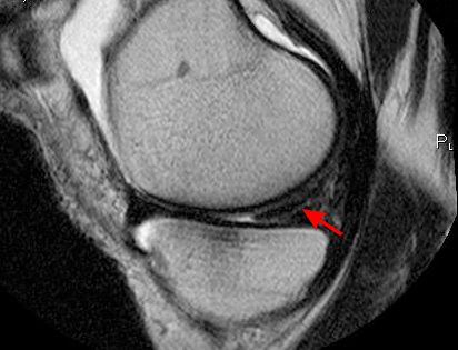
Result
signal changes within meniscus on T1 and T2 images; sagittal views: anterior and posterior meniscal tears; coronal view: far medial and far lateral meniscal tears
x-ray
Test
Anterior-posterior x-ray, lateral knee x-ray, 45° posteroanterior flexion, and skyline views are indicated in patients with a history of arthritis and chronic, long-standing meniscal tears.[3]
If the leg is malaligned, a long-leg hip to ankle film should also be obtained.
Fairbanks' changes are due to abnormal force concentration in the joint consequent to the loss of protection provided by the meniscus.
Result
arthritis and/or chronic tears: flattening of femoral condyle, narrowing of joint space, osteophyte formation, subchondral sclerosis (Fairbanks' changes)
Investigations to consider
arthroscopy
Test
Although usually performed to repair or resect the torn meniscus, arthroscopy can be used to confirm diagnosis.
Result
torn meniscus
Use of this content is subject to our disclaimer