Images and videos
Images


Assessment of maculopapular rash
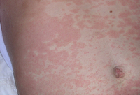
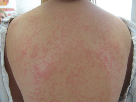
Drug eruption due to phenytoin
Photograph courtesy of Brian L. Swick
See this image in context in the following section/s:

Assessment of maculopapular rash
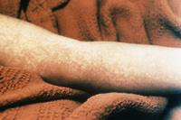
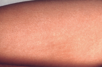
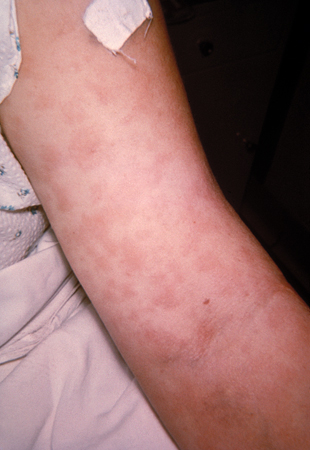
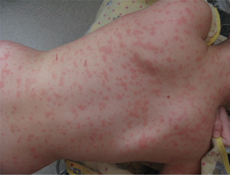
Scarlet fever rash on the forearm due to group A Streptococcus bacteria
Courtesy of the CDC Public Health Image Library
See this image in context in the following section/s:
Assessment of maculopapular rash
During a punch biopsy, local anaesthetic is instilled by injection into the surrounding skin. With the skin stretched perpendicular to normal relaxation lines, a disposable skin biopsy punch with a round stainless steel blade (at least 3 mm or 4 mm in diameter is recommended) is applied perpendicular to the skin. Pressure is applied with a rotating action until a ‘give’ is felt as the blade pierces through the subcutaneous tissue. The cylindrical core of tissue is removed and placed in a labelled sample pot containing a suitable fixative solution
Created by the BMJ Knowledge Centre
See this image in context in the following section/s:

Assessment of maculopapular rash
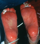
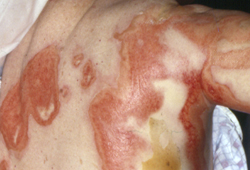
Stevens-Johnson syndrome: epidermal loss on soles of feet
From the personal collection of Dr A. Kowal-Vern
See this image in context in the following section/s:

Assessment of maculopapular rash
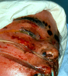
Toxic epidermal necrolysis with epidermal loss, ocular involvement, and ecthyma gangrenosum
From the personal collection of Dr A. Kowal-Vern
See this image in context in the following section/s:
Assessment of maculopapular rash
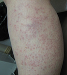
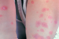
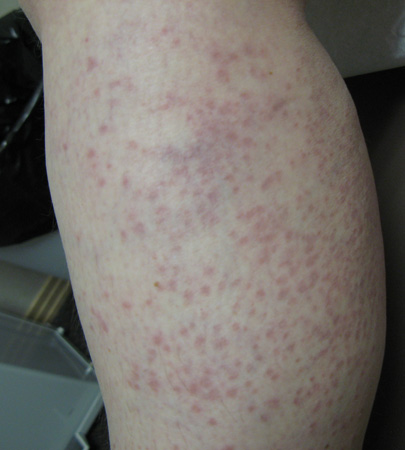
Viral exanthem presenting as a maculopapular eruption. Note the erythematous papules are larger than macules on the leg of a young adult
Photograph courtesy of Hobart W. Walling
See this image in context in the following section/s:
Assessment of maculopapular rash
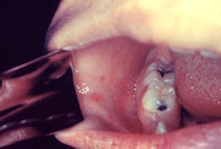
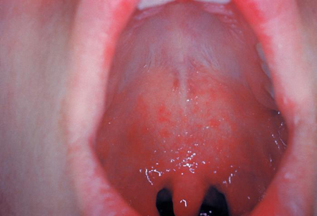
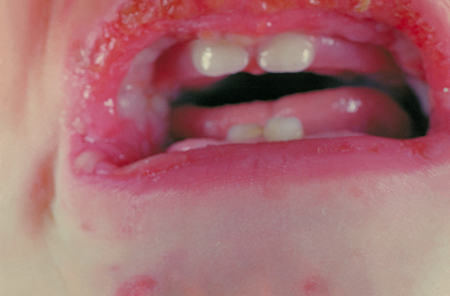
Koplik's spots
Centers for Disease Control and Prevention
See this image in context in the following section/s:

Assessment of maculopapular rash
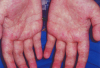
Syphilis presenting with a generalised rash
Courtesy of the CDC Public Health Image Library
See this image in context in the following section/s:
Assessment of maculopapular rash
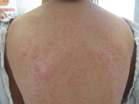
Viral exanthem presenting as a maculopapular eruption. Note urticarial appearance of non-scaling erythematous macules and papules on the trunk of this older child
Photograph courtesy of Hobart W. Walling
See this image in context in the following section/s:
Assessment of maculopapular rash
Stevens-Johnson syndrome: targetoid lesions and epidermal loss
From the personal collection of Dr A. Kowal-Vern
See this image in context in the following section/s:

Assessment of maculopapular rash
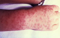
Characteristic spotted rash of Rocky Mountain spotted fever
Courtesy of the CDC Public Health Image Library
See this image in context in the following section/s:
Assessment of maculopapular rash
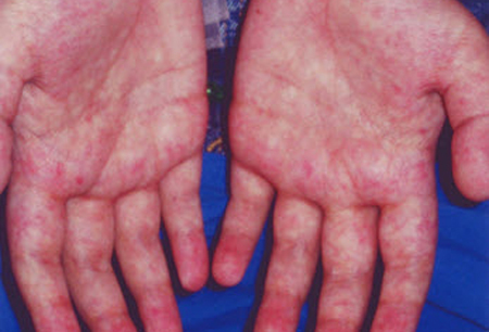
Hypersensitivity rash due to penicillin
CDC Public Health Image Library
See this image in context in the following section/s:

Assessment of maculopapular rash
Morbilliform rash (resembling measles) resulting from toxic shock syndrome
Courtesy of the CDC Public Health Image Library
See this image in context in the following section/s:
Assessment of maculopapular rash
Erythema multiforme with perioral ulceration
Courtesy of the CDC Public Health Image Library
See this image in context in the following section/s:
Assessment of maculopapular rash
Acute graft-versus-host disease of the skin (grade I)
Courtesy of Dr John Levine, Professor, Blood and Marrow Transplantation Program, University of MIchigan; used with permission
See this image in context in the following section/s:
Assessment of maculopapular rash
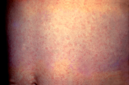
Rubella presenting as a generalised abdominal rash
Courtesy of the CDC Public Health Image Library
See this image in context in the following section/s:

Assessment of maculopapular rash
Erythema multiforme with target lesions
Courtesy of the CDC Public Health Image Library
See this image in context in the following section/s:

Assessment of maculopapular rash
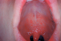
Koplik's spots
Centers for Disease Control and Prevention
See this image in context in the following section/s:
Assessment of maculopapular rash
Erythema infectiosum (Fifth disease) in a 10-year-old girl
From the personal teaching collection of Hobart W. Walling
See this image in context in the following section/s:
Use of this content is subject to our disclaimer