Aetiology
The trachea and oesophagus arise from the common foregut, initially starting as a single tube, then dividing into two distinct tracheal structures. The trachea has lung buds at the caudal end of the primitive trachea. Separation starts during the fourth week of gestation, and failure of normal division can result in a spectrum of defects including atresias, fistula formation, and tracheal clefts. Three major theories explain how the respiratory and digestive foreguts arise from a single progenitor, which might inform the morphological origin of oesphageal atresia (OA) and tracheo-oesophageal fistula (TOF).[7] While none of the theories can be proven without live imaging of the developing embryo, evidence favours the development of the separate respiratory and oesophageal foregut from septation of the common foregut.[8]
OA/TOF often occurs as an isolated defect and has a low twin-twin concordance rate, which led past investigators to conclude the condition was primarily non-genetic. However, due to OA/TOF’s association with other congenital anomalies in up 50% of cases, this failure in organogenesis may be secondary to single gene mutations or chromosomal disorders. A number of candidate genes and their affiliated molecular networks have been implicated in oesophagal and pulmonary dysmorphogenesis, indicating the complex signalling necessary for the development of these structures.[9]
Pathophysiology
When OA is present, the infant is unable to swallow any liquid, including his or her own secretions. The infant cannot drink until either the atresia is repaired, or the stomach is accessed via the anterior abdominal wall. If a fistula is present, the connection between the airway and the alimentary tract must be ligated and divided. In type C, this connection can cause gaseous distension of the stomach and small bowel on x-ray. In rare cases, there is an associated intestinal atresia, which can lead to over-expansion and rupture of the stomach. Gastric contents can also reflux back through the fistula and cause aspiration, resulting in a chemical and bacterial pneumonitis.[10]
Motility of the oesophagus is always affected, with the distal segment having the most marked disordered peristalsis.
There is also a lower resting pressure of the lower oesophageal sphincter resulting in a higher incidence of GORD. Investigators have determined this dysmotility is related to maldevelopment or malfunction of the enteric nervous system that forms the dual autonomic plexuses common to the entire human gut.[11][12]
Classification
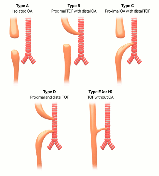
Gross classification: the surgery of infancy and childhood, 1954[1]
Type A
Pure atresia (4% to 7%)
Type B
Proximal fistula with distal atresia (1%)
Type C
Proximal atresia with distal fistula (85% to 90%)
Type D
Proximal and distal fistula (3%)
Type E
H-type fistula (2% to 3%)
Tracheo-oesophageal fistula without oesophageal atresia
[Figure caption and citation for the preceding image starts]: Types of tracheo-oesophageal fistula/oesophageal atresia based on Gross classificationCreated by the BMJ Knowledge Centre [Citation ends].

Use of this content is subject to our disclaimer