Investigations
1st investigations to order
clinical examination
Test
A complete ophthalmological examination should be performed.
Visual acuity may be difficult to ascertain in children. It is important to check for a relative afferent pupillary defect, as visual loss due to increased orbital pressure is a real concern in these patients.[1][9][10][11]
Result
orbital signs such as decreased vision, a relative afferent pupillary defect, dysmotility, chemosis, and proptosis may be present in orbital cellulitis; eyelid oedema and erythema are commonly seen in both pre-septal and orbital cellulitis.
CT sinus and orbits with contrast medium
Test
The mainstay diagnostic method for suspected cases of orbital cellulitis.[12][24]
Peri-orbital cellulitis: inflammation of peri-orbital tissues anterior to the orbital septum; absence of orbital inflammation.[Figure caption and citation for the preceding image starts]: Axial CT post contrast: opacified ethmoid and frontoethmoidal recess on the left sideFrom the personal collections of H. Jane Kim, MD, and Robert Kersten, MD, UCSF; used with permission [Citation ends].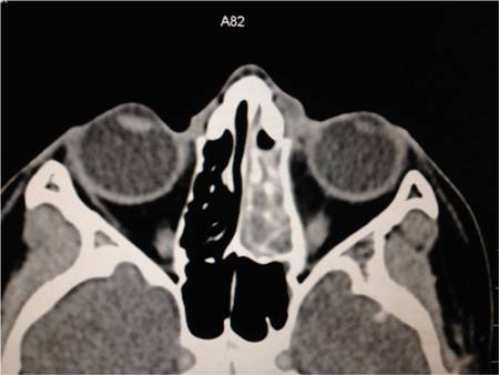 [Figure caption and citation for the preceding image starts]: Coronal CT post contrast: opacified ethmoid and frontoethmoidal recess on the left sideFrom the personal collections of H. Jane Kim, MD, and Robert Kersten, MD, UCSF; used with permission [Citation ends].
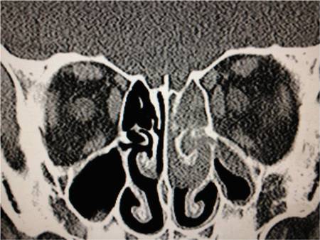
Orbital cellulitis: inflammation of orbital tissues located deep to the septum.
Result
inflammation of peri-orbital or orbital tissue; sub-periosteal abscess (commonly superomedial or inferomedial); sinusitis
Investigations to consider
blood culture
Test
Sample may be obtained before starting antibiotic therapy.
Results are likely to be negative, more so in adults than in children.[21][23] Positive culture rates are between 0% and 33%.[22]
Organisms may include Staphylococcus species,Streptococcus species, and anaerobes. Haemophilus influenzae is rarely cultured in immunised individuals.
Result
positive smear, positive growth
microbiology swab (conjunctiva, nasopharynx, external wounds)
Test
May be collected from conjunctiva, nasopharynx (especially if a fungal infection is suspected), external wounds, or tissue obtained during surgery, as necessary.
Result
positive smear, positive growth
MRI head and orbits with contrast medium
Test
May be performed in patients demonstrating neurological deficit.[12]
Useful in soft tissue disease evaluation, but its application is limited due to availability and the need for sedation in children.
Result
evidence of intracranial abscess or cavernous sinus thrombosis
Orbital ultrasonography
Test
Can differentiate between pre-septal and orbital infection.[25]
May have advantages for some patients as it can be performed at the bedside and does not require sedation; however, it is unable to adequately assess associated sinus or intracranial involvement.[19][26]
Result
inflammation of peri-orbital or orbital tissue; sub-periosteal abscess
Use of this content is subject to our disclaimer