Investigations
1st investigations to order
ophthalmological evaluation; computerised visual-field examination
Test
Performed in all patients with craniopharyngioma with suprasellar extension and chiasmal compression.
Result
may reveal unilateral or bitemporal hemianopsia
MRI brain (contrast-enhanced)
Test
Most sensitive and specific imaging modality. [Figure caption and citation for the preceding image starts]: Craniopharyngioma: coronal post-contrast MRIFrom the collection of Marc C. Chamberlain [Citation ends].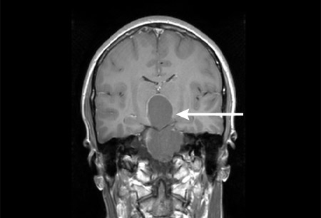 [Figure caption and citation for the preceding image starts]: Craniopharyngioma: axial post-contrast MRIFrom the collection of Marc C. Chamberlain [Citation ends].
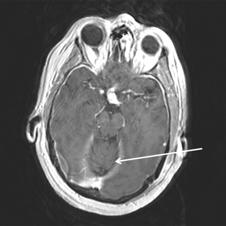 [Figure caption and citation for the preceding image starts]: Craniopharyngioma: sagittal post-contrast MRIFrom the collection of Marc C. Chamberlain [Citation ends].
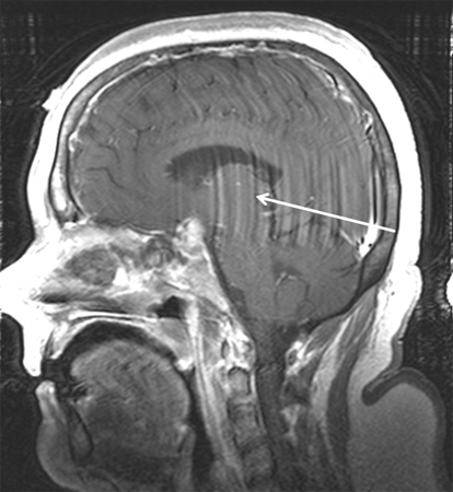 [Figure caption and citation for the preceding image starts]: Craniopharyngioma: sagittal post-contrast MRIFrom the collection of Marc C. Chamberlain [Citation ends].
Allows the clinician to define the size, location, and relationship of the tumour to surrounding structures; to determine the surgical approach; to assess the extent of resection; and to plan radiotherapy.
Result
variable; T1-weighted imaging may show hyperintensity secondary to high protein content in cystic component; contrast-enhanced sequences show enhancement of the solid component and cyst wall in mixed solid-cystic lesions; T2-weighted imaging and fluid-attenuated inversion recovery (FLAIR) show heterogeneous signal in the solid components and cyst hyperintensity; calcification is hypointense on T2-weighted imaging
CT brain (contrast-enhanced)
Test
Used when MRI is not available or if there are contraindications to MRI.
Of value in demonstrating tumour location in relation to the sella, and in assessing response to treatment during follow-up.
Result
frequent tumour calcification (90% children; 70% adults); mixed cystic and solid mass with enhancement of the solid component and cyst wall
serum prolactin
Test
Increased secretion is due to tumour compression of the pituitary stalk.
Result
variable; commonly elevated
serum growth hormone (GH)
Test
Used to diagnose GH deficiency.
Result
variable; commonly depressed
serum insulin-like growth factor 1 (IGF-1)
Test
Used to diagnose growth hormone deficiency.
Result
variable; commonly depressed
serum insulin-like growth factor binding protein-3 (IGFBP-3)
Test
Used to diagnose growth hormone deficiency.
Result
variable; commonly depressed
provocative growth hormone (GH) tests
Test
Provocative agents (e.g., levodopa, insulin, glucagon) are given to stimulate the pituitary to release GH.
Used to diagnose GH deficiency; may be required if other screening tests (GH, insulin-like growth factor 1, insulin-like growth factor binding protein-3) are equivocal.
Result
variable; commonly may show failure to induce GH
serum luteinising hormone
Test
Used to diagnose gonadotrophin hormone deficiency.
Result
variable; commonly depressed
serum follicle-stimulating hormone
Test
Used to diagnose gonadotrophin hormone deficiency.
Result
variable; commonly depressed
morning serum testosterone
Test
Used to diagnose gonadotrophin hormone deficiency in men.
Blood should be drawn between 8 a.m. and 9 a.m.
Result
variable; commonly depressed
serum thyroid-stimulating hormone and T3/T4
Test
Used to diagnose thyroid hormone deficiency.
Result
variable; commonly depressed
morning serum cortisol and adrenocorticotrophic hormone (ACTH)
Test
Used to diagnose adrenal insufficiency.
Blood should be drawn between 8 a.m. and 9 a.m., when cortisol levels peak.
It is important to realise that diabetes insipidus cannot occur in the presence of chronic low mineralocorticoids; administration of corticosteroids can unmask low vasopressin and result in the onset of severe diabetes insipidus.
Result
variable; commonly depressed in association with non-elevated levels of ACTH
serum electrolytes
Test
Used to diagnose diabetes insipidus.
Result
variable; hypernatraemia
urine and serum osmolality
Test
Used to diagnose diabetes insipidus.
Result
variable; commonly elevated plasma osmolality
urine specific gravity
Test
Used to diagnose diabetes insipidus.
Result
variable; commonly low
plain x-rays for bone age
Test
Used to diagnose growth hormone deficiency.
Result
often show a delayed bone age in children
Investigations to consider
tissue histology
Test
Allows for definitive diagnosis following surgical biopsy/resection with pathological analysis of tumour tissue. [Figure caption and citation for the preceding image starts]: Craniopharyngioma: adamantinous histology (low power) with complex arrangements of epithelium, cysts, and gliotic brainFrom the collection of Marc C. Chamberlain [Citation ends].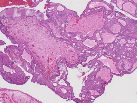 [Figure caption and citation for the preceding image starts]: Craniopharyngioma: adamantinous histology (medium power) with epithelial ribbons showing reticular areas and nodules of keratinFrom the collection of Marc C. Chamberlain [Citation ends].
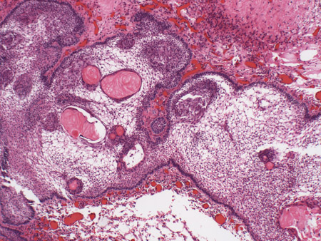 [Figure caption and citation for the preceding image starts]: Craniopharyngioma: adamantinous histology (high power) with basal-aligned columnar cells, stellate reticulum, and epithelial keratinisationFrom the collection of Marc C. Chamberlain [Citation ends].
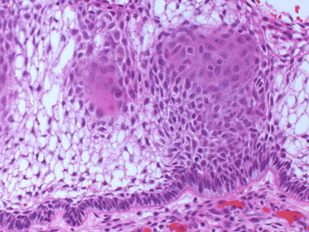
Result
adamantinous/squamous epithelial tumour; calcification
Use of this content is subject to our disclaimer