Images and videos
Images


Craniopharyngioma
Craniopharyngioma: coronal post-contrast MRI
From the collection of Marc C. Chamberlain
See this image in context in the following section/s:
Craniopharyngioma
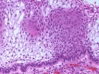
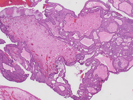
Craniopharyngioma: adamantinous histology (low power) with complex arrangements of epithelium, cysts, and gliotic brain
From the collection of Marc C. Chamberlain
See this image in context in the following section/s:
Craniopharyngioma
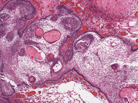
Craniopharyngioma: adamantinous histology (medium power) with epithelial ribbons showing reticular areas and nodules of keratin
From the collection of Marc C. Chamberlain
See this image in context in the following section/s:

Craniopharyngioma
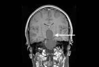
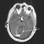
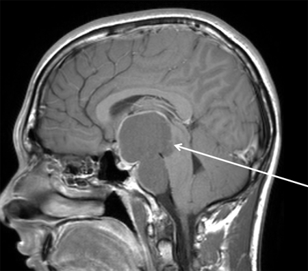
Craniopharyngioma: axial post-contrast MRI
From the collection of Marc C. Chamberlain
See this image in context in the following section/s:
Craniopharyngioma
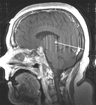
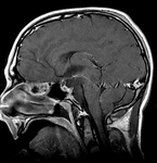
Craniopharyngioma: sagittal post-contrast MRI
From the collection of Marc C. Chamberlain
See this image in context in the following section/s:

Craniopharyngioma
Craniopharyngioma (postoperative): sagittal post-contrast MRI
From the collection of Marc C. Chamberlain
See this image in context in the following section/s:

Craniopharyngioma
Craniopharyngioma: adamantinous histology (high power) with basal-aligned columnar cells, stellate reticulum, and epithelial keratinisation
From the collection of Marc C. Chamberlain
See this image in context in the following section/s:

Craniopharyngioma
Craniopharyngioma: sagittal post-contrast MRI
From the collection of Marc C. Chamberlain
See this image in context in the following section/s:
Use of this content is subject to our disclaimer