Approach
History and physical examination
The most common tool in diagnosis of erythema infectiosum is the clinical appearance of the classic 'slapped cheek' rash with a lacy, reticular exanthem on the extremities and torso. There may or may not be a history of low-grade fever and non-specific influenza or respiratory symptoms occurring prior to onset of the exanthem.
Patients may develop arthralgia/arthritis, particularly of the small joints of the hands, wrists, knees or ankles, but these joint symptoms are usually self-limiting. When presented with this clinical scenario in an otherwise healthy individual, no other diagnostic investigations are required.
Patients who are immunocompromised do not form immune complexes, so they do not present initially with classic erythema infectiosum. This patient population includes patients with HIV, those receiving chemotherapy or immunosuppression following transplant, or patients with congenital immunodeficiencies.[2] Persistent parvovirus B19 infection may occur, with resultant severe anaemia due to chronic red cell aplasia.[Figure caption and citation for the preceding image starts]: Typical erythematous 'slapped cheeks' of erythema infectiosum.From the collection of Gary A. Dyer, MD; used with permission [Citation ends].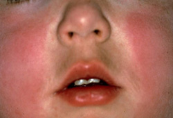 [Figure caption and citation for the preceding image starts]: Lacy, reticular, erythematous eruption of erythema infectiosum on an upper extremity.From the collection of Gary A. Dyer, MD; used with permission [Citation ends].
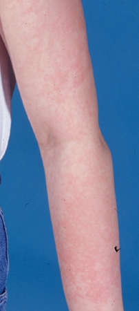
Full blood count (FBC)
An FBC and reticulocyte count is recommended in pregnant patients and patients with known immunosuppression or conditions involving increased red blood cell turnover/destruction (e.g., hereditary spherocytosis, sickle cell disease, thalassaemia, iron deficiency anaemia), to examine for anaemia. Consultation with haematology/oncology consultants is recommended in the setting of a transient aplastic crisis (a decrease in haemoglobin levels with absent reticulocytes during acute infection).
Serology
Serological studies are rarely necessary except in the setting of complicated parvovirus B19 infection (e.g., investigation into the cause of severe fetal anaemia or the cause of aplastic crises).[2] Serology may be helpful in a pregnant woman presenting with known exposure to the virus and/or the typical exanthem or arthralgias, routine serological screening is not recommended in pregnancy, as the risk of seroconversion is low, and fetal outcomes vary.[25] If serological testing is performed, immunoglobulin M (IgM) positivity indicates acute infection, regardless of immunoglobulin G (IgG) status, and requires monitoring for potential fetal infection. In cases where both IgM and IgG are negative, the woman is susceptible to infection and should undergo repeat serological testing after 4 weeks to assess for seroconversion.[26] Women who are IgM negative and IgG positive have immunity to parvovirus B19 and are protected from infection.
IgM antibodies are present in most cases of erythema infectiosum at the time of diagnosis and may develop in the days after onset of aplastic crisis. IgM antibodies are detectable for several months after acute infections. Maternal IgM may not be present during the onset of fetal hydrops, although IgM can be detected in umbilical cord blood and DNA studies (of cord blood or amniotic fluid) can detect the virus.[27]
Patients who are immunocompromised may not be able to eradicate the infection due to inadequate levels of IgM. They may remain infectious, but test negative to IgM serology. Alternative assays, including nucleic acid hybridisation and polymerase chain reaction assays may be helpful in confirming suspected diagnosis. As viral DNA may be detected for months after the viremic phase, a positive result may not be evidence of acute infection.
Use of this content is subject to our disclaimer